Extracranial to Intracranial Bypass in Down Syndrome Related Moyamoya and Improved Seizure Controls
Abstract number :
3.348
Submission category :
9. Surgery / 9C. All Ages
Year :
2018
Submission ID :
499023
Source :
www.aesnet.org
Presentation date :
12/3/2018 1:55:12 PM
Published date :
Nov 5, 2018, 18:00 PM
Authors :
Michael Doherty, Swedish Epilepsy Center; Stephen Monteith, Swedish Neuroscience Institute; Sarah Garson, Swedish Primary Care Green Lake; Nicole Warner, Swedish Epilepsy Center; Bart Keogh, Radia Imaging; and Ryder Gwinn, Swedish Epilepsy Center
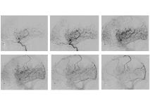
Rationale: Epilepsy can be a common side effect of progressive intracranial vascular narrowings seen in moyamoya. In this study we look at outcomes on epilepsy control in a patient with Down syndrome and moyamoya who underwent an extracranial to intracranial bypass procedure. Methods: A 27-year-old patient with Down syndrome and epilepsy presented with subacute worsening of seizure control. Seizures types varied and in general began as focal onset hyperkinetic events with progression to bilateral tonic clonic or atonic drops. In addition a second type of focal onset impaired awareness events with behavior and motor arrest with non-responsivity were evident. Fatigue, participation and atonic drops with convulsive activity worsened (two per week) and were leading to injuries and problems walking. Quality of life became markedly worse necessitating gait assistance, caregiver support on transfers, decreased verbal participation and heightened supervision. Medications that were once effective-clobazam, lamotrigine, levetiracetam- became less so . Workup showed intracranial arterial narrowings mainly in bilateral MCA territories consistent with moyamoya, lateral left internal carotid angiogram views are included in Figure 1. Perfusion abnormalities over MCA territories were evident and had progressed over time suggesting a dynamic process that paralleled her clinical worsening (figure 2, column B shows increased perfusion times in red over anterior MCA territories bilaterally). Staged bilateral revascularization procedures using a superior temporal to middle temporal artery bypass were offered. Results: Left indirect superior temporal artery to left middle cerebral artery bypass was initially performed, with excellent postoperative course. Three months after the initial surgery the right superior temporal artery to left middle cerebral artery bypass was done, again with a straightforward postoperative course. Outcome improvements included: less fatigue, decreased anti epileptic drug requirements, initiation of conversations (which she had stopped doing in childhood) and an absence of seizures at 6 months post second bypass. Repeated imaging showed improvements in perfusion, with the last vertical labelled pair "C" showing increases in perfusion over MCA territories globally. Conclusions: In a Down syndrome patient with progressive perfusion abnormalities due to moyamoya and resultant hard-to-treat epilepsy, revascularization of bilateral MCA territories via superior temporal artery bypass improved quality of life, seizure control, verbal output, fatigue, gait and medication burdens. Funding: None

.tmb-.jpg?Culture=en&sfvrsn=2bd35645_0)