A DISTINCT MALFORMATION OF CORTICAL DEVELOPMENT IN FRONTAL LOBE OF CHILDREN WITH FOCAL EPILEPSY
Abstract number :
1.110
Submission category :
Year :
2005
Submission ID :
5161
Source :
www.aesnet.org
Presentation date :
12/3/2005 12:00:00 AM
Published date :
Dec 2, 2005, 06:00 AM
Authors :
Prakash Kotagal, William Bingaman, Paul Ruggieri, and Guyan Wu
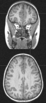
We report two children with intractable focal epilepsy and a distinctive malformation of cortical development involving their frontal lobes. Two children 6 and 13 years old at the time of epilepsy surgery, began having seizures from the age of 4 and 1.5 years respectively. Seizures were complex motor or versive seizures followed by secondary generalization. Other than borderline low IQ or moderate mental retardation with ADHD and behavior problems, there were no focal neurologic deficits. After scalp video-EEG evaluation, PET, ictal/interictal SPECT and neuropsychological evaluation, they were studied with subdural grid electrodes combined with depth electrodes in one patient. Interictal scalp EEG showed multifocal spikes in the left hemisphere, not only in the left frontal lobe but also the temporal and parietal regions as well as generalized spikes in the younger child. Scalp ictal EEG was lateralized to the left hemisphere in one; in the other, it showed left fronto-polar onset with spread to the left occipital region. MRI scans were reported as normal in one patient and in the other a deep superior frontal sulcus was apparent.
FDG-PET showed striking hypometabolism in the left frontal lobe corresponding to the cortex lining the anomalous sulcus. Ictal SPECT showed localized hyperperfusion in the same region as the PET abnormality in one; in the other an ictal injection was not obtained but localized hypoperfusion was noted on the baseline study. Both children were studied with subdural grid electrodes; one child also had depth electrodes inserted into the sulcus. Ictal EEG onset was very diffuse in the younger child; in the other it was localized to the superior and mid frontal regions. Both underwent focal resections (guided by PET and SPECT findings in the younger child) that spared the anatomical Broca[apos]s area. They have been entirely seizure free for 6 years and 1 year respectively. One child came off AEDs successfully. Histopathology showed mild to moderate cortical dyplasia. Striking frontal hypometabolism seen on FDG-PET scans of children with intractable epilepsy, should lead to closer examination for the presence of an anomalous deep superior frontal sulcus with surrounding dysplastic cortex. Focal resection of the involved cortex may lead to an excellent seizure outcome despite poorly localized EEG findings.[figure1]
