A Novel Approach for Accurate Automatic Stereotactic EEG Electrode Identification and Grouping from Post-Implantation Scans
Abstract number :
1.128
Submission category :
2. Translational Research / 2E. Other
Year :
2018
Submission ID :
502642
Source :
www.aesnet.org
Presentation date :
12/1/2018 6:00:00 PM
Published date :
Nov 5, 2018, 18:00 PM
Authors :
Noam Peled, MGH/Harvard; Taha Gholipour, Brigham and Women'S Hospital; Angelique Paulk, MGH/Harvard; Sydney Cash, MGH/Harvard; Darin Dougherty, MGH/Harvard; Emad Eskandar, MGH/Harvard; Matti Hamalainen, MGH/Harvard; and Steven M. Stufflebeam, MGH/Harvard
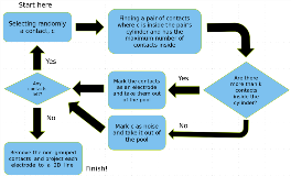
Rationale: Pre-surgical work up for resective surgery or neuromodulation device insertion in some patients with drug-resistant epilepsy involves invasive electrographic recording to better delineate the seizure onset zone and/or to map eloquent cortices. Understanding the exact location of each electrode contact in relation to cortical and sub-cortical landmarks is crucial during the interpretation of the recording. Currently, many clinicians and use a manual labeling approach, and some algorithms are available for grids and strips; however, there are no published methods for labeling stereotactic EEG (sEEG) automatically. Methods: Using computer modeling, we developed a novel algorithm for detecting and labeling depth electrodes. We used 1mm post-SEEG implantation CT from 13 patients (mean=36.64 years old, range 19 to 58; 7 females) with drug-resistant epilepsy to validate our method. Briefly, we assume a cylinder-shaped model for depth electrodes without a priori knowledge of the number of electrodes or contacts, but with a preset minimum acceptable distance between two contacts. Local maxima optimization is used to first detect the contacts on the CT, which makes the method sensitive for finding all the contacts, but may generate false detections. Next, the Random Sample Consensus (RANSAC) approach is used to group the contacts into the different electrodes, and therefore to resolve any false detections. Through an iterative process, contacts are placed in cylinder-shaped models with 3mm diameter, and those not fitting are labeled as noise. The CT is then registered to pre-implantation 1mm isotopic MPRAGE MRI. Everything outside the dura is removed using FreeSurfer reconstruction, but a soft inside/outside function is used to include the contacts that seem outside of the brain. The contacts in each electrode are projected to a 2D line and then used in our multi-modal visualization tool (MMVT) over the 3D model of each patient’s brain for manual adjustments. Results: Without a priori number of electrodes and contacts, the algorithm detected all the electrodes in 13 patients (13±3 electrodes and 156±52 contacts per patient). 95% (1924 out of 2025) of the contacts were correctly identified and grouped into their electrodes, and 69 contacts were falsely detected (3.4%). The hours-long task of manual identification was reduced to minutes per patient. After visualization in MMVT, the human reviewer could interactively resolve any remaining ambiguities. Conclusions: We present a new method for automatic detection and grouping of SEEG electrode contacts. We show high sensitivity and specificity, comparable to time-consuming manual detection in our sample patient group. A combination with a multimodal visualization tool allows fast and reliable data that can improve interpretation of intracranial recording and pre-surgical planning. Future studies will focus on clinical validation using larger samples. Funding: Partially funded by DARPA (W911NF-14-2-0045, SUBNETS Program); NCRR (S10RR014978) and NIH (S10RR031599, R01-NS069696, 5R01-NS060918, U01MH093765).

.tmb-.png?Culture=en&sfvrsn=638b82af_0)