A Proposed Model for Investigating Functional Connectivity and Glutamate/GABA Concentrations as Biomarkers of Epilepsy Using 7T MRI
Abstract number :
2.198
Submission category :
5. Neuro Imaging / 5B. Functional Imaging
Year :
2018
Submission ID :
501730
Source :
www.aesnet.org
Presentation date :
12/2/2018 4:04:48 PM
Published date :
Nov 5, 2018, 18:00 PM
Authors :
Ofer M. Gonen, The Royal Melbourne Hospital; Patrick Kwan, University of Melbourne, The Royal Melbourne Hospital; Terence J. O'Brien, Monash University; Patricia M. Desmond, The Royal Melbourne Hospital; Bradford A. Moffat, University of Melbourne; and El
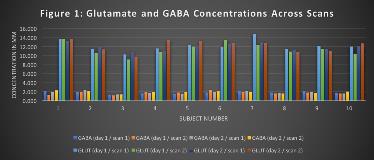
Rationale: Functional connectivity (FC) of the default mode network (DMN) has been shown to be reduced in patients with epilepsy compared with healthy controls. Furthermore, evidence from several studies suggests that in certain types of epilepsy the FC differences correlate with duration of epilepsy, response to treatment, and seizure focus laterality. The posterior cingulate cortex (PCC)/precuneus is the main node of the DMN. Previous studies investigating the relationship between the concentrations of the main excitatory and inhibitory cortical neurotransmitters, glutamate and gamma-aminobutyric acid (GABA), and FC of the PCC/precuneus, have shown conflicting results with field strengths up to 3 Tesla. The advent of 7-Tesla MRI technology enables accurate quantification of glutamate and GABA in the PCC/Precuneus via Magnetic Resonance Spectroscopy (MRS), which can be correlated with FC measures. Methods: Ten healthy right-handed volunteers ranging in age from 23-68 underwent 7-Tesla MRI scans on two different days. Resting-state fMRI was acquired on the first day. MRS of a single 8 mL voxel, located in the PCC/precuneus, was acquired via Stimulated Echo Acquisition Mode (STEAM) twice on the first day and twice on the second day, with a break for re-positioning in between. All scans were preceded by high-resolution anatomical scans for localisation and co-registration purposes. LCModel was used to calculate glutamate and GABA concentrations in the chosen voxel, which were normalised according to grey matter percentage to correct for partial volume effects. FC was calculated between the PCC/Precuneus and the other main nodes of the DMN (medial prefrontal cortices and lateral parietal cortices) via the CONN toolbox. Correlation coefficients were converted to normally distributed scores using Fisher’s transformation. For correlation and regression analysis the MRS results immediately acquired after resting-state fMRI were chosen. Results: The mean coefficients of variation within sessions of glutamate and GABA were 10.30% and 5.72%, respectively; and between sessions 5.21% and 3.42%, respectively. Both mean FC of the PCC/precuneus to the other DMN nodes and the glutamate/GABA ratio correlated with age (R=0.78, p=0.007; R=0.71, p=0.023, respectively). Multiple regression demonstrated no correlation between the glutamate/GABA ratio and the FC of the PCC/precuneus after adjustment for age (adjusted R2=0.51, p=0.899). Conclusions: Acquiring 7-Tesla MRS of PCC/precuneus metabolites is feasible and reproducible. Both glutamate/GABA ratio and FC of the PCC/precuneus correlate with age, but are otherwise unrelated and can be combined in a multimodal pipeline to investigate differences between types of epilepsy. Funding: The Royal Melbourne Hospital Neuroscience Foundation. The 7T scanner and BAM are supported by the National Imaging Facility via the Australian Government NCRIS program.

.tmb-.jpg?Culture=en&sfvrsn=dabf9bf3_0)