Abnormal Hippocampal Structure and Function in Patients With Juvenile Myoclonic Epilepsy and Their Unaffected Siblings: A Magnetic Resonance Imaging Study
Abstract number :
2.166
Submission category :
5. Neuro Imaging / 5A. Structural Imaging
Year :
2018
Submission ID :
500873
Source :
www.aesnet.org
Presentation date :
12/2/2018 4:04:48 PM
Published date :
Nov 5, 2018, 18:00 PM
Authors :
Lorenzo Caciagli, University College London; Britta Wandschneider, University College London; Fenglai Xiao, University College London; Christian Vollmar, Ludwig-Maximilians-Universitaet; Maria Centeno, University College London; Karin Trimmel, University
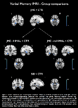
Rationale: Juvenile myoclonic epilepsy (JME) is the most frequent genetic generalised epilepsy syndrome, characterised by a complex polygenetic aetiology. Structural and functional MRI studies demonstrated mesial or lateral frontal cortical derangements and impaired fronto-cortico-subcortical connectivity in patients and their unaffected siblings. The presence of hippocampal abnormalities and associated memory deficits is controversial, and functional MRI (fMRI) studies in JME have not tested hippocampal activation. Here, we implemented multi-modal MRI and neuropsychological data to investigate structure and function of the mesiotemporal lobe in patients with JME and their unaffected siblings. Methods: We recruited 37 patients with juvenile myoclonic epilepsy, 16 unaffected siblings and 20 healthy controls, comparable for age, gender, handedness and hemispheric dominance as assessed with language lateralisation indices. Automated hippocampal volumetry was complemented by qualitative and quantitative morphological criteria to detect hippocampal malrotation (HIMAL; Figure 1), assumed to represent a neurodevelopmental marker. Neuropsychological measures of verbal and visuospatial learning and an event-related verbal and visual memory fMRI paradigm addressed mesiotemporal function. Results: We detected a reduction of left hippocampal volume (5-8%) in patients and their siblings compared with controls (p < 0.01). Unilateral or bilateral hippocampal malrotation was identified in 51% of patients and 50% of siblings, against 15% of controls (p < 0.05). Logistic regression analysis, including gender and handedness, identified group allocation as a highly significant predictor of hippocampal malrotation (p < 0.01). For bilateral hippocampi, quantitative markers of verticalization had significantly larger values in patients and siblings compared with controls (MANOVA, p < 0.05). In the patient subgroup, there was no relationship between structural measures and earlier disease onset or poorer seizure control. No overt impairment of verbal and visual memory was identified with memory tests. Functional mapping highlighted atypical patterns of hippocampal activation, pointing to dysfunction of verbal encoding in patients and their siblings (Figure 2). Subgroup analyses indicated distinct profiles of hypoactivation along the hippocampal long axis in JME with and without malrotation (Figure 2), and a more prominent role of the left posterior hippocampus for visual memory in patients with malrotation (all p < 0.05, FWE-corrected). Linear models across the entire study cohort indicated significant associations between quantitative morphological markers of hippocampal positioning and hippocampal activation for verbal items (all p < 0.05, FWE-corrected). Conclusions: Our findings document hippocampal morphometric abnormalities implicating volume, shape and positioning in patients with JME and their siblings, which relate to reorganisation of function, and likely stem from an underlying neurodevelopmental mechanism with expression during the prenatal stage. Co-segregation of imaging patterns in patients and their siblings is suggestive of genetic imaging phenotypes, independent of disease activity. The mesiotemporal lobe is identified as neural system affected by genetic variants predisposing to JME. Funding: Wellcome Trust (Project Grant No 079474); Henry Smith Charity (Ref. 20133416); Brain Research UK (personal fellowship to LC); Medical Research Council; Wolfson Trust; Epilepsy Society; National Institute for Health Research University College London Hospitals Biomedical Research Centre; Deutsche Forschungsgemeinschaft

.tmb-.png?Culture=en&sfvrsn=6b4e5a05_0)