AFTER DISCHARGE (AD) STIMULATION ON SIMULTANEOUS SCALP-EEG AND INTRACRANIAL EEG (ECoG) AND SPIKE SOURCE RECONSTRUCTION
Abstract number :
2.173
Submission category :
Year :
2003
Submission ID :
3970
Source :
www.aesnet.org
Presentation date :
12/6/2003 12:00:00 AM
Published date :
Dec 1, 2003, 06:00 AM
Authors :
Dinah Thyerlei, Tatiana Maleeva, Adam N. Mamelak, Felix I. Darvas, William W. Sutherling MEG Unit, Huntington Medical Research Institutes, Pasadena, CA; Epilepsy and Brain Mapping, Pasadena, CA; Signal and Image Processing Institute, University of Souther
Cortical stimulation of intracranial grid electrodes is used to identify functionally relevant areas before brain surgery. We recorded grid electrode stimulation and simultaneous scalp-EEG. Stimulating an intracranial electrode causes a local high current directly under the contact, which may cause AD and thereby provides a known current source location at the patient[rsquo]s cortex. We reconstructed the spike source from both ECoG and scalp-EEG and compared the results to the true location of the stimulated grid contact.
Patients undergoing pre-surgical evaluation with intracranial electrodes (ECoG) were measured. All contacts were stimulated to determine the AD threshold and identify functionally important areas. Stimulation was performed with symmetric biphasic pulses for 0.3 ms, with a 50 Hz frequency and 2-5 s trains. The current intensity was increased every 20 s starting at 1 mA up to 15 mA or until AD occurred. Simultaneous 64-channel scalp-EEG was recorded with a 200 Hz sampling rate. Three landmarks and all electrode positions were digitized. The intracranial grid electrode positions were taken from a post-surgical CT. The AD spikes seen simultaneously in grid and scalp electrodes were used to fit single moving dipoles +/- 50 ms around the peak. Localization accuracy was compared using a three-shell sphere and a BEM model for the scalp-EEG data and a single sphere for the ECoG data (Curry 4.6).
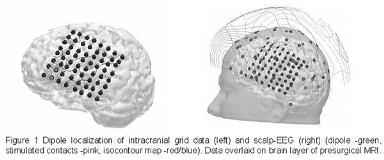
Fitting a single moving dipole to AD-spikes using ECoG and a single sphere leads to a distance of 3.6 [plusmn] 1.5mm from the dipole localization to the center of the two stimulated contacts (s. fig.1 left). The same spikes measured on simultaneous scalp-EEG have an error of 31.2 [plusmn] 10.7mm using a three shell-sphere (s. fig.1 right). Results for a more realistic head model (BEM) depend critically on the anatomical accuracy of the specific model used.
Co-registration of intracranial and scalp-EEG allows for a direct evaluation of surface based source reconstructions. Due to many postsurgical alterations (e. g. skull changes, edema, the non-conductive silicon layer of the electrode grid) simple head models might not yield satisfactory results, but even with a three-shell sphere model a localization accuracy of 30mm can be achieved. Due to postsurgical inhomogeneities inside the brain and skull holes a BEM model does not necessarily perform better. Using a sophisticated FEM might improve the results.[figure1]
[Supported by: NIH grants NINDS RO1-NS20806, NCRR S10RR13276, NIHM MH53213, and the Norris, Zeilstra and Bacon Foundations.]
