Age-Specific Pediatric Head Atlases for Forward Electrical Modeling
Abstract number :
1.181
Submission category :
3. Neurophysiology / 3G. Computational Analysis & Modeling of EEG
Year :
2018
Submission ID :
482308
Source :
www.aesnet.org
Presentation date :
12/1/2018 6:00:00 PM
Published date :
Nov 5, 2018, 18:00 PM
Authors :
Sergei Turovets, Philips Neuro; Kyle Morgan, Philips Neuro; David Hammond, Oregon Institute of Technology; Jidong Hou, Philips Neuro; Amanda McCutcheon, Philips Neuro; Kai Li, Philips Neuro; Ha Eun Kim, Philips Neuro; Kirk Smith, University of Arkansas fo
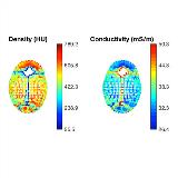
Rationale: Accurate cortical source estimation from magneto- and electroencephalography (MEG/EEG) ideally relies on subject specific forward head models that enable computation of lead fields to the scalp. With the widespread use of MRI and CT availability anatomically realistic head models can be used routinely, however there is still a lack of age specific models for pediatric populations, where CT and MRI scanning is inappropriate for shear research use. While there is an increasing number of open-source platforms that offer atlas-based realistic head models, most of them are limited to the adult population. In this paper, we describe the construction and verification of pediatric age-appropriate atlases for EEG. Methods: Retrospective pediatric CT and MRI volumes were acquired in public domains and by data mining the clinical image repository of the Washington University Health System in St. Louis, MO. Individual head models were created by co-registering subject matched MRI and CT. Age-group atlases were built by nonlinear warping of the publicly available probabilistic non-linear- average MRI templates to our age matched full head CTs, and subsequent segmentation with in-house segmentation software. The quality of segmentation was validated by visual slice-by-slice inspection and manual correction. The nonlinear warping of brain templates into CT crania was validated by matching CSF layers to the average CSF thickness at several landmarks. The models are complimented by inhomogeneous skull conductivity maps based on the Hounsfield units CT intensity patterns (Fig. 1) and anisotropic brain conductivity by co-registering the average implanted brains with the average pediatric brain DTIs (Fig.2). The performance of atlases was evaluated by source localization of scalp somatosensory evoked potentials (SEPs) induced by a finger piezo-tapper. Results: Pediatric volumes of 66 matched MRI/CT of good quality were identified from which 40 individual head models were derived using in-house segmentation software to serve as a look up table in source localization. We have also created four age group atlases for i) neonates; ii) infants (0-2 y); ii) preschoolers (3-8 y) and iv) school children (9-17 y) by fusing the UNC 0 and 1 y, the MNI 4-8 y and 10-14 y average MRIs to our neonatal, 1 y, 5.5 y and 13.5 y CT templates. We included four 8 y subjects in prospective SEP studies. The comparative source localization results showed that the optimal anatomical face validation occurred when the age appropriate atlas was used. In addition, 12 age-specific nonlinear average templates of T1, T2 MRI and DTI volumes have been created using the ANTs software platform and the NIH pediatric MRI data base. Conclusions: The age group pediatric atlases are useful assets for EEG source localization. They are freely provided to the scientific community for research use (www.pedeheadmod.net). In addition to EEG/MEG, they are relevant to optical imaging modeling problems as well and can be used to define common coordinate systems for analysis of multi-modal imaging data. Funding: This research was supported by NINDS Grant R43NS67726 and NIMH Grant R44MH106421.

.tmb-.jpg?Culture=en&sfvrsn=e6e247de_0)