Analysis of Phase-Amplitude Coupling Between Fast and Slow Oscillations in Scalp EEG in Patient With Epileptic Spasms Without MRI Abnormality
Abstract number :
2.095
Submission category :
3. Neurophysiology / 3G. Computational Analysis & Modeling of EEG
Year :
2018
Submission ID :
501734
Source :
www.aesnet.org
Presentation date :
12/2/2018 4:04:48 PM
Published date :
Nov 5, 2018, 18:00 PM
Authors :
Hidenori Sugano, Juntendo University; Madoka Nakajima, Juntendo University; Yasushi Iimura, Juntendo University; Takumi Mitsuhashi, Juntendo University; Kaito Kawamura, Juntendo University; Takuma Higo, Juntendo University; and Hajime Arai, Juntendo Unive
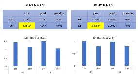
Rationale: The mechanism of epileptic spasms (ES) have still under discussed. Recently, the analysis of modulation index (MI) that reflecting the degree of coupling between high frequency oscillations (HFO) and slow waves in the electrocorticography was reported as providing information to find out the epileptic foci in patients with ES. Epileptic areas showed HFO coupled with 3-4Hz slow wave in previous studies. However, the reported method is invasive and the next step for us is to detect meaningful MI even in scalp EEG. Because detecting HFO in scalp EEG was difficult, we tried to analyze the phase-amplitude coupling between the gamma and slow oscillations in a patient with ES. Methods: We analyzed long term scalp EEG of 7-year-old boy with 6-years history of ES. He received 4 times ACTH therapy before the corpus callosotomy (CCS). His seizure frequency was a few series of ES a day. EEG showed spike & wave complex in multiple foci. We performed total CCS and achieved improvement of his ES in number and severity. We analyzed IEDs for five minutes and five times at 500 Hz of sampling rate for MI. Electrodes of each hemisphere was calculated using EEGLAB Toolbox PACTv.0.17 in MATLAB. We measured MIs between amplitude of 3-4 Hz oscillation and the following two bands of gamma oscillations; low gamma oscillations (LGO) ranging from 30 to 50Hz, and high gamma oscillations (HGO) ranging from 50 to 80Hz. We compared MIs in each hemisphere before and after the CCS. Results: Visual inspection of EEG presented no change of IED characteristics even after CCS. Before CCS, MIs in each hemisphere did not show any differences in both MIs of LGO-slow and HGO-slow oscillations. Even post CCS, MIs of those between each hemisphere did not result any difference. However, changes of MIs in left hemisphere reduced after CCS with statistically significant (p<0.01). On the other hand, those in right hemisphere did not show any reduction. Patient's seizure semiology changed ES with left side dominance after CCS. Conclusions: Analyzing the phase-amplitude coupling in scalp EEG demonstrated the dynamic changes of the epileptogenicity in ES after CCS. Continuing high MI between gamma and 3-4Hz slow oscillations could mean pathological epileptogenicity. Corpus callosum plays the crucial role to enhance the symptomatic and electrical severities in ES. Funding: None
