Anatomical Understanding of Subtotal Hemispherotomy Using Cadaveric Brain, 3D-Simulation Models, and Intraoperative Photographs
Abstract number :
2.316
Submission category :
9. Surgery / 9B. Pediatrics
Year :
2018
Submission ID :
480065
Source :
www.aesnet.org
Presentation date :
12/2/2018 4:04:48 PM
Published date :
Nov 5, 2018, 18:00 PM
Authors :
Takehiro Uda, Osaka City University Graduate School of Medicine; Ryoko Umaba, Osaka City University Graduate School of Medicine; Kosuke Nakajo, Osaka City University Graduate School of Medicine; Yuta Tanoue, Osaka City University Graduate School of Medici
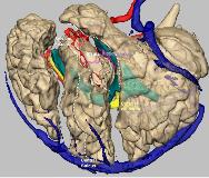
Rationale: Hemispherotomy that preserves the primary motor and sensory areas is called subtotal hemispherotomy. It is a combined procedure involving temporo-parieto-occipital disconnection and frontal disconnection, but because of the narrow and deep surgical window and complex anatomical relationships, it may result in insufficient disconnection, with continuation of seizures. In the present study, we attempted to understand this surgery using cadaveric brains, three-dimensional (3D) simulation models, and intraoperative photographs. Methods: A formalin-fixed cadaveric brain was dissected to show each step of this surgery. For 3D simulation, 3D models of several brain structures were reconstructed from preoperative imaging, and the surgery was simulated. Intraoperative photographs were taken from a case of a 12-year-old girl with epileptic spasm. Results: Fronto-temporo-parietal craniotomy over the entire part of the Sylvian fissure (SF), postcentral sulcus, and precentral sulcus was performed. The SF was dissected widely. The insular cortex was exposed, and corticotomy was made on the inferior periinsular sulcus (IPS) until the inferior horn of the lateral ventricle (LV) was opened. The corticotomy was extended proximally, and the limen insulae was dissected. The uncal gyrus was dissected, and the dissection was extended posteriorly to the inferior choroidal point. The corticotomy along the IPS was extended distally to the atrium of the LV. The fornix was dissected, and another corticotomy was made along the postcentral sulcus. At the bottom of the corticotomy, the body of the LV was opened. The corticotomy was then extended laterally to the distal part of the SF and connected with the corticotomy along the IPS. Next, the corticotomy was also extended medially to the interhemispheric fissure (IH) and the corpus callosum (CC) along the falx cerebri. The splenium of the CC was dissected from the wall of the LV. In these steps, the temporo-parieto-occipital lobe and the limbic system were disconnected. Then, another corticotomy was made along the precentral sulcus. At the bottom of the corticotomy, the body of the LV was opened. The corticotomy was extended laterally and connected to the SF. Along the SF, the corticotomy was extended to the frontal base. The orbital gyrus and rectal gyrus were disconnected until the IH. The corticotomy along the precentral sulcus was also extended medially to the IH and to the CC along the falx cerebri. From the LV, the body, genu, and rostrum of the CC were dissected. The subcallosal area was dissected, and the dissection plane connected with the lateral corticotomy. With these steps, the frontal lobe was disconnected. Conclusions: Combined use of cadaveric brains, 3D simulation models, and intraoperative photographs facilitates a clearer anatomical understanding of subtotal hemispherotomy. Funding: None
