Asymmetry in Resting State Functional MRI Connectivity Networks for Characterization of Temporal Lobe Epilepsy
Abstract number :
2.189
Submission category :
5. Neuro Imaging / 5B. Functional Imaging
Year :
2018
Submission ID :
501186
Source :
www.aesnet.org
Presentation date :
12/2/2018 4:04:48 PM
Published date :
Nov 5, 2018, 18:00 PM
Authors :
Taha Gholipour, Brigham and Women'S Hospital; Douglas Greve, Martinos Center for Biomedical Imaging, Massachusetts General Hospital; Jong Woo Lee, Brigham and Women's Hospital; and Steven M. Stufflebeam, Martinos Center for Biomedical Imaging, Massachuset
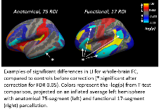
Rationale: Temporal lobe epilepsy (TLE) is considered a brain network disease. Different measures have been used to demonstrate alterations of the resting state functional connectivity (FC) networks in TLE. Since FC values are vary across subjects, here we propose to use a laterality index (LI) derived from the raw FC values for comparing predefined region of interests (ROIs) to identify and characterize TLE. Methods: Data from 42 people with left TLE and 26 controls were used for voxel-wise whole-brain analysis in FreeSurfer Functional Analysis Stream). The degree of FC was defined as the number of connections with a correlation threshold higher than 0.5 to: a) all other voxels of the brain, b) voxels limited to a surface-based neighborhood (local FC), and c) voxels outside that neighborhood (distant FC). The LI was calculated using three parcellation methods to represent asymmetry in pre-defined ROIs: 35- and 75-segment anatomical parcellation ROIs available in FreeSurfer, and ROIs from a 17-network parcellation method. The coefficient of variation for LI was compared to raw FC values from which LI is calculated to determine variability. LI values from TLE were compared to controls using student's T-test, adjusted for a false discovery rate of 0.05. Results: Compared to raw FC, LI showed lower variability of distribution across subjects. The LI values were visualized by projection of significance probability (p-value) to a surface ROI map. A decrease in the LI of the inferior temporal region was demonstrated (LI -0.03 vs. 0.16 for controls, 75-segment method, p<0.0005). The ROI named functional network-17 (involving parts of the lateral temporal cortex and functionally connected distant cortices) showed a similar decrease in asymmetry (LI 0.06 vs. 0.14 for controls, p<0.005). The hippocampus ROI also showed significant decrease in asymmetry (p<0.05). A difference was noted between the pattern of local and distant FC asymmetry in each region. Conclusions: Asymmetry in FC provides a lower inter-subject variability for investigating FC. Using LI in pre-defined anatomical and functional ROIs to healthy controls, a decreased asymmetry in inferior temporal FC was shown. A functional network involving parts of lateral temporal cortex in TLE shows a similar decrease. Additionally, the pattern of FC asymmetry appears to be different when looking at local versus distant FC. We propose that future studies focus on specific asymmetry patterns, perhaps using novel machine learning methods to improve the diagnosis and treatment of TLE. Funding: This work was supported by the AES/TS Alliance Research Training Fellowship for Clinicians to TG.
