Changes in Interictal and Ictal EEG Patterns Following Complete Corpus Callosotomy
Abstract number :
1.357
Submission category :
9. Surgery / 9B. Pediatrics
Year :
2018
Submission ID :
507135
Source :
www.aesnet.org
Presentation date :
12/1/2018 6:00:00 PM
Published date :
Nov 5, 2018, 18:00 PM
Authors :
Maite La Vega-Talbott, Mount Sinai Health System; Malgosia Kokoszka, Mount Sinai Health System; Dina Bolden, Mount Sinai Health System; Patricia McGoldrick, Icahn School of Medicine at Mount Sinai; Karen Lob, Washington University in, St. Louis; Hillary R
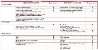
Rationale: We examined pre- and post-surgery EEG characteristics of patients undergoing complete corpus callosotomy (CC) in order to better characterize possible electrographic outcomes of the procedure. Methods: Scalp video EEG (VEEG) recordings from before and after callosotomy were reviewed by 2 providers not originally involved in the patients’ care. The last VEEG performed prior to CC, the first performed after, and the most recent available study were analyzed. Additional post-CC VEEGs were also reviewed if the first study after CC did not capture any ictal events. Results: Fifty patients were identified who underwent complete CC over a period of 15 years (June 2003 – May 2018). Pre- and at least one post-CC VEEG were readily available for 40 of those patients (28M, 12F, mean age at surgery 11 ± 8 years, median age 9, range 1-44 years). Background EEG findings, seizure semiology, and ictal EEG features from before and after CC are summarized in Table 1. Prior to callosotomy, 39 patients had generalized onset seizures with interictal generalized epileptiform abnormalities, and all 40 had generalized background slowing. Twenty-three patients had 1 seizure type, 15 had 2, and 2 patients had 3 seizure types captured, with atonic semiology being the predominant seizure type in nearly half (19) of the patients, followed by tonic and myoclonic seizures (10 cases of each). Following callosotomy, 22 patients had only 1 and 13 more had 2 seizure types captured, and atonic semiology was found in only 9 cases. Further, in 10 cases ictal EEG lateralized with focal motor seizures or (new onset) behavioral arrest. Three patients experienced remarkable electrographic improvement following callosotomy – e.g. developed a normal posterior rhythm of 8 Hz alpha in the awake state and had fewer epileptiform discharges with less background activity. Of the 20 patients who had at least 3 VEEGs reviewed for this study, 8 had no seizures captured on the most recent one. Finally, 21, or more than half of patients, had no changes in seizure characteristics. Conclusions: This study illustrates a range of possible electrographic changes following complete corpus callosotomy, from no change in EEG pattern to improved background, to lateralization and focal onset. As new surgical modalities such as responsive neurostimulation become available for epilepsy, a detailed re-examination of outcomes after CC will help to determine optimal approaches and the utility of CC for an individual patient. Funding: The study was supported by the Department of Neurosurgery, Mount Sinai Health System.
