Clinical Utility of Magnetic and Electric Source Imaging in the Localization of Interictal Spikes Prior to Pediatric Epilepsy Surgery
Abstract number :
2.058
Submission category :
3. Neurophysiology / 3D. MEG
Year :
2018
Submission ID :
502320
Source :
www.aesnet.org
Presentation date :
12/2/2018 4:04:48 PM
Published date :
Nov 5, 2018, 18:00 PM
Authors :
Michel Alhilani, Boston Children's Hospital, Harvard Medical School; Eleonora Tamilia, Boston Children's Hospital, Harvard Medical School; Melissa Tsuboyama, Boston Children's Hospital, Harvard Medical School; Jurriaan M. Peters, Harvard Medical School, B
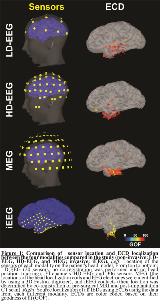
Rationale: Magnetic and electric source imaging (MSI and ESI) facilitate the estimation of the epileptogenic zone (EZ) in patients with medically refractory epilepsy (MRE) by enabling the localization of the irritative zone (IZ). Several studies demonstrated the utility of MSI and ESI in the IZ localization by comparing their findings with intracranial electroencephalography (iEEG) data. Few studies correlated both ESI and MSI to iEEG in the same cohort of patients, but none performed ESI on iEEG to compare the same quantitative localization measures. Our goal is to assess the clinical utility of magnetoencephalography (MEG) and scalp electroencephalography (EEG) in estimating the IZ prior to pediatric epilepsy surgery by: (i) calculating the localization error (ELoc) and precision of the IZ localized non-invasively (using MSI and ESI on MEG and scalp EEG) with respect to the ground-truth IZ localized invasively (using ESI on iEEG) and (ii) correlating the resection of the noninvasively-localized IZ with surgical outcome. Methods: We studied 24 children with MRE who underwent surgery after iEEG. We analyzed 10-15 minutes of interictal data from: (i) simultaneous MEG (306 sensors) and high-density (HD) EEG, 70 channels); (ii) low-density (LD) EEG (20 channels), and (iii) iEEG (116±40 subdural and/or depth contacts) (Fig 1). IEDs were identified on each modality by two reviewers (only IEDs marked by both were further analyzed). We co-registered the presurgical magnetic resonance imaging (MRI) with the post-implantation computerized tomography to determine the iEEG electrode location, and with postsurgical MRI to determine the resected volume. We localized the activity at the IED peak using an Equivalent Current Dipole (ECD). The patient’s presurgical MRI was used to build a realistic head model and a Boundary Element Model (BEM) consisting of skin, skull and brain tissue (we used three BEM layers for MEG, HD- and LD-EEG, and only brain tissue for iEEG). We calculated the minimum Euclidean distance (Dmin) of each ECD from any iEEG contact: ECDs with a Dmin >30 mm were classified as outside-coverage. For ECDs within coverage, we calculated the ELoc, as Dmin from any iEEG ECD: ECDs with ELoc =15 mm were classified as concordant with iEEG. The precision of each modality was estimated as percentage of concordant ECDs. Finally, we classified each ECD as resected (Dmin from resection =15 mm) or not resected. Results: MEG showed the lowest percentage of outside-coverage ECDs (MEG: 7%; HD-EEG: 19%; LD-EEG: 31%; ?2: p<0.001), and the highest precision (MEG: 59%; HD-EEG: 46%; LD-EEG: 28%; ?2: p<0.001, Fig 2). MEG-ECDs showed the lowest ELoc(12 mm), followed by HD-EEG (17 mm) and LD-EEG (23 mm, ANOVA: p<0.001). Finally, for iEEG, MEG and HD-EEG, the resection of the ECDs was associated with a good outcome (p<0.001), while no association was found for LD-EEG. Conclusions: MSI and ESI are valuable tools for the presurgical mapping of interictal activity in children with MRE. Our data indicates that MEG localizes the IZ more precisely than HD- and LD-EEG. The resection of the IZ localized using iEEG, MEG or HD-EEG was associated with good outcome emphasizing the clinical utility of MSI and ESI in the estimation of the EZ. Funding: No funding

.tmb-.jpg?Culture=en&sfvrsn=9c042843_0)