CORRELATION OF MAGNETIC RESONANCE SPECTROSCOPIC IMAGING AND INTRACRANIAL EEG LOCALIZATION OF SEIZURES
Abstract number :
1.124
Submission category :
Year :
2005
Submission ID :
5176
Source :
www.aesnet.org
Presentation date :
12/3/2005 12:00:00 AM
Published date :
Dec 2, 2005, 06:00 AM
Authors :
1Edward Scharff, 1Hitten Zaveri, 1,2Xenophon Papademetris, 1Hal Blumenfeld, 1Robert B. Duckrow, 3Hoby P. Hetherington, 1Susan S. Spencer, 1Dennis D. Spencer, 1,2<
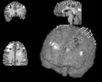
Currently, the [ldquo]gold standard[rdquo] for the localization of seizures in intractable cases of epilepsy is intracranial electroencephalography (icEEG). Though highly effective, icEEG requires that electrode leads be surgically implanted, and the cost and complexity associated with this procedure presents a barrier to treatment that might be mitigated by less expensive and invasive methods. Magnetic resonance spectroscopic imaging (MRSI) is a new, noninvasive tool that has proven to be effective for lateralizing seizures in temporal lobe epilepsy. The goal of this study is to establish correlation between the results of traditional icEEG methods and measurements obtained using proton (1H) MRSI. We identified 9 patients with intractable epilepsy who had 1H-MRSI taken prior to intracranial electrode implantation. Single and multislice 1H MRSI was performed on a 4 Tesla Varian MR system. Data was obtained using a previously published pulse sequence with 8mm x 8mm x 10mm resolution. For each spectroscopy voxel, we calculated the difference between the measured ratio of creatine to N-acetyl-aspartate and an expected ratio based on the grey matter content of that voxel in a group of normal controls. These values were used to determine lateralization of seizures.Subsequently, the patients underwent intracranial electroencephalography and seizure localization and lateralization was determined by ictal EEG recordings. The intracranial EEG electrode locations were co-registered with the MRSI data using software developed at Yale. For each patient, we compared the clinical lateralization made using icEEG methods with MRSI lateralization results. The figure demonstrates the co-registration of MRSI, MRI and intracranial electrodes on a subject. Voxels where the NAA/Cr ratio on MRS was significantly different from controls are highlighted. We found that icEEG and MRSI lateralization was concordant in all 9 subjects. It is apparent from these results that MRSI is a reliable method of lateralizing seizures. MRSI is capable of much higher spatial resolution. Further analysis correlating quantitative measures of icEEG with MRSI data on a voxel by voxel basis are underway. These studies are needed to determine whether noninvasive MRSI can be used to localize seizures more precisely.[figure1] (Supported by NIH - NINDS and R01EB000473.)
