Cortical Thickness in Childhood Left Focal Epilepsy: Thinning Beyond the Seizure Focus
Abstract number :
3.247
Submission category :
5. Neuro Imaging / 5A. Structural Imaging
Year :
2018
Submission ID :
507343
Source :
www.aesnet.org
Presentation date :
12/3/2018 1:55:12 PM
Published date :
Nov 5, 2018, 18:00 PM
Authors :
Emanuel Boutzoukas, Children's National Medical Center; Jason Crutcher, National Institutes of Health; Eduardo Somoza, Howard University; Leigh Sepeta, Children's National Hospital System/NINDS NIH; Xiaozhen You, Children's National Hospital System/NINDS
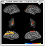
Rationale: Patients with localization-related epilepsy, including adults and children, demonstrate widespread decreased cortical thickness (CT) that extends beyond the ipsilateral epileptogenic zone (Widjaja et al., 2012). Pediatric epilepsy study samples have been relatively small and heterogeneous with regard to seizure type and hemisphere of seizure focus. Therefore, we examined if CT differed between children with left hemisphere focal epilepsy (LHE) and age-matched typically developing (TD) peers. Within clusters of group differences, we then examined if CT was associated with duration of epilepsy or neuropsychological functioning, including executive functioning (EF) that has not previously been examined in pediatric epilepsy. Methods: 35 children with LHE (mean age: 10.46 years) and 35 TD controls (mean age: 10.47 years) completed neuropsychological testing and 3T MR imaging. All participants had normal MRI. All surface-based morphometric processing and analyses were conducted using FreeSurfer (FS) 6.0, including FS’ statistical tool, Qdec. Data were smoothed to improve intersubject averaging with a 15 mm full width half maximum Gaussian kernel. To correct for multiple comparisons, a Monte-Carlo simulation was implemented with a cluster-forming threshold set to p<0.05. Bivariate correlations were conducted to examine the relationship between CT and duration of epilepsy. Neuropsychological measures correlated with CT results included general intelligence (WASI PIQ and VIQ), and executive functioning (parent reported Behavior Rating Inventory of Executive Functioning (BRIEF) Behavioral Regulation (BRI) and MetaCognition Indices (MCI)). Results: CT was significantly decreased for children with LHE compared to TD children across 6 clusters bilaterally (2 Left; 4 Right; Figure 1) including left frontoparietal-cingulate cortex, an area at the parieto-occipital junction bilaterally, and right supramarginal, paracentral, and superior frontal gyri. Seizure duration was not significantly correlated with CT.Greater PIQ was associated with greater CT in the left frontoparietal-cingulate region (r=.332, p<0.001), left and right parieto-occipital junction (left r =.260, p=0.04; right r=.280, p=0.03), and right supramarginal gyrus (r=.284, p=0.02). The right parieto-occipital junction was also positively correlated with VIQ (r=.320, p=0.01). More parent reported problems of EF were associated with decreased CT in the left frontoparietal-cingulate region (BRIEF MCI r=-.250, p=0.04) and the right superior frontal gyrus (BRIEF BRI r=-.269, p=0.03; BRIEF MCI r=-.248, p<0.05). Conclusions: Children with LHE displayed widespread cortical thinning, extending beyond the seizure focus location and to the contralateral hemisphere. This thinning was associated with lower IQ and greater everyday executive dysfunction. Seizure duration was not associated with CT suggesting that perhaps these structural differences are evident early on. If these differences precede epilepsy onset, CT may be a useful biomarker for risk of neurological dysfunction. Funding: NINDS 1K23NS065121-01A2

.tmb-.png?Culture=en&sfvrsn=5d03c86f_0)