Diffuse Cortical Thinning in BECTS Is Evident by 2 Years After Diagnosis
Abstract number :
1.255
Submission category :
5. Neuro Imaging / 5A. Structural Imaging
Year :
2018
Submission ID :
500666
Source :
www.aesnet.org
Presentation date :
12/1/2018 6:00:00 PM
Published date :
Nov 5, 2018, 18:00 PM
Authors :
Sally M. Stoyell, Massachusetts General Hospital; Mckenna Parnes, Massachusetts General Hospital; Daniel Y. Song, Massachusetts General Hospital; Lauren M. Ostrowski, Massachusetts General Hospital; Emily L. Thorn, Massachusetts General Hospital; Grace Xi
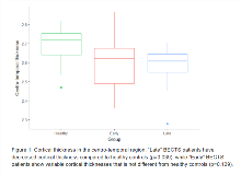
Rationale: Benign epilepsy with centrotemporal spikes (BECTS) is a common childhood epilepsy syndrome, characterized by a transient period of seizures that co-occurs with a dramatic period of cortical development. Children with BECTS display a stereotyped EEG pattern with spikes in the centrotemporal region. Previous studies in BECTS have shown inconsistent, patchy regions of increased (Kim et al. 2015, Overvliet et al. 2013), decreased (Overvliet et al. 2013) or unchanged cortical thickness (Garcia-Ramos et al. 2015) overlapping with centrotemporal regions. We hypothesized that there is a transient period of cortical thinning in the centrotemporal cortex evident later in disease. We use an a priori approach to evaluate cortical thickness in centrotemporal regions between children with BECTS and healthy controls (HC) based on disease duration. We also perform post-hoc analyses to evaluate the spatial specificity of these findings. Methods: High resolution T1-weighted 3T MRI scans were collected in children with BECTS (n=23) and HC (n=18). Cortical thickness measures were computed using Freesurfer v5.3. The centrotemporal area and regional cortical thickness measures were calculated for each subject. The centrotemporal area in BECTS was defined here by the precentral, postcentral and superior temporal gyri labels from the Freesurfer Desikan-Killiany atlas. Cortical thickness differences were compared between BECTS patients based on duration of disease (“Early”, within 2 years of first seizure; “Late”, more than 2 years from first seizure), and HC. Longitudinal data was evaluated in a subset of patients (n=4). Age was included as a covariate in all analyses. Results: We found a significant difference in cortical thickness in the centrotemporal gyri between groups (ANOVA, p=0.027). “Late” BECTS patients differed significantly from HC (Tukey HSD test, estimate=-0.122mm; SE=0.048; adjusted p=0.039), while the difference between “Early” BECTS patients and HC was not significant (adjusted p=0.129). We found similar results when looking across brain regions (frontal, temporal, parietal, occipital, insula; MANOVAs: p<0.001 "Late" vs HC; p0.6 “Early” vs HC). Each brain region evaluated was significantly thinner in “Late” BECTS compared to HC, (p< 0.05 for all tests, one-way ANOVA, FDR corrected). In the subset of BECTS patients with longitudinal data, preliminary data supports a possible relationship between disease duration and rate of cortical thinning in the centrotemporal region (n=4; p=0.057), where BECTS children showed a transient period of accelerated thinning corresponding to disease onset. Conclusions: Our results suggest that children with BECTS have transient, diffuse thinning of the cortex that is evident by 2 years after diagnosis. Attention to disease duration may explain the prior apparently contradictory results in the literature. Future work should be done to examine the functional significance of these findings.References:Garcia-Ramos et al. Epilepsia. 2015;56:1615-1622.Kim et al. Seizure. 2015;27:40-46.Overvliet et al. Neuroimage. 2013;2:434-439. Funding: NINDS K23NS092923

.tmb-.png?Culture=en&sfvrsn=e99cce79_0)