Disrupted Basal Ganglia Pathways in Temporal Lobe Epilepsy With Focal to Bilateral Seizures
Abstract number :
3.249
Submission category :
5. Neuro Imaging / 5B. Functional Imaging
Year :
2018
Submission ID :
502634
Source :
www.aesnet.org
Presentation date :
12/3/2018 1:55:12 PM
Published date :
Nov 5, 2018, 18:00 PM
Authors :
Xiaosong He, Thomas Jefferson University; Ganne Chaitanya, Thomas Jefferson University; Burcu Asma, Thomas Jefferson University; Ellen Eline, Thomas Jefferson University; Noah Sideman, Thomas Jefferson University; Hela Saidi, Thomas Jefferson University;
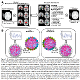
Rationale: The prevalence of focal to bilateral tonic-clonic seizures (FBTCS) is associated with severe morbidity, higher risk of SUDEP and poor treatment outcomes1. While little is known about the critical circuitry involved in FBTCS, some basal ganglia (BG) structures such as thalamus have been implied in such process2. Hence, we offered a network science perspective to articulate the functional organization of the BG associated with FBTCS in temporal lobe epilepsy (TLE). Methods: Five-min resting-state fMRI data were collected from 72 TLE patients, who were classified into 3 groups (24 patients each, L/R TLE=12/12) based on their seizure history: with focal seizures only (F), with remote FBTCS (rFBTCS, no episode for at least a year), and with current FBTCS (cFBTCS). We first applied masked ICA to a group of high-resolution healthy control data to segment the striatum, globus pallidus (GP), and thalamus into 20 functional parcels per hemisphere (Fig1A). We then extracted the parcel time series from our clinical fMRI data. Considering the unilateral nature of the BG pathways, we calculated a functional connectivity (FC) matrix for each hemisphere. The 2 matrices for RTLE were switched left-to-right to maintain access to ictal vs. non-ictal side. We then estimated the modular identity of each parcel from each FC matrix, and compared it with its anatomical identity to calculate the integration index (the probability a parcel is assigned to the same module with parcels from a different region), as a measure of inter-regional communication (Fig1B). Results: The 3 patient groups did not differ in age, gender, handedness, or other major clinical characteristics. Multivariate ANOVA revealed a significant group difference in the integration between ictal-sided GP and thalamus (F=8.33, PFDR=0.003) (Fig2A). We further broke down the striatum (putamen, caudate) and GP (external, internal), and found a significant group difference in the integration between the ictal-sided GP internal (GPi) and thalamus (F=9.85, PFDR=0.004) (Fig2B), where cFBTCS patients showed reduced integration compared to the other two groups. A Jonckheere-Terpstra test showed a significant ordinal effect (TJT=4.08, PFDR<0.001), indicating a gradual reduction of integration here (cFBTCS

.tmb-.png?Culture=en&sfvrsn=aa58852a_0)