EPILEPTIFORM LOCALIZATION FROM SIMULTANEOUS EEG-FMRI RECORDINGS
Abstract number :
1.244
Submission category :
Year :
2003
Submission ID :
3846
Source :
www.aesnet.org
Presentation date :
12/6/2003 12:00:00 AM
Published date :
Dec 1, 2003, 06:00 AM
Authors :
Michiro Negishi, Hal Blumenfeld, Dennis D. Spencer, R. Todd Constable Diagnostic Radiology, Yale University, New Haven, CT; Neurology, Yale University, New Haven, CT; Neurobiology, Yale University, New Haven, CT; Neurosurgery, Yale University, New Haven,
Combination of electroencephalography (EEG) and functional magnetic resonance imaging (fMRI) for epileptiform localization has been an active area of research (EEG triggerd fMRI: e.g. Neurology 1996, 47, 89-93, EEG-fMRI simultaneous recording: e.g. Proc. ISMRM 1998, 168). However, a better understanding of the difference between EEG and fMRI signals is necessary to fully benefit from the both modalities. The current paper presents a comparison of EEG source estimation and fMRI spatial parameter mapping from data that are simultaneously collected from epileptic patients.
Five runs of 10-15 minute EEG-fMRI recordings are conducted for each epileptic patient. The fMRI parameters are:1.5T (GE) echo-planer, TR=3 Sec., TE=50 mSec., 64 x 64 x18 voxels. The EEG data is sampled at 500 Hz (Neuroscan 22 to 32 channels system). Epileptiforms from each subject are averaged to obtain a time-course of a spike-wave pair and are subjected to source localization using the MUSIC algorithm (IEEE Trans. Antenas. Propagat. 1986, AP-34, 276-280). From standardized fMRI volumes, eight volumes including one that contains the onset of each spike are selected and a spatial parametric map (SPM99, Hum Brain Mapp 1995, 2, 189-210) is computed using epileptiform periods as regressors.
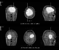
EEG source localization maps and an fMRI spatial parameter map from a patient are shown below as an example. The EEG source estimation on the spike part of the epileptiforms (Fig. 1(a)) results in an activation in the left dorsal frontal cortex, whereas that on the wave part (b) results in the right ventral frontal cortex. The SPM result (Fig. 2) shows an activity in the left parietal lobe ([x, y, z] = [-16,-52,36], left precuneus) as the only statistically significant activity (p=0.026). There is a weaker left activity closer to the EEG spike localization. There is also a weak right activity in the parameter map, which is inferior compared to the EEG wave localization. Precision of EEG wave localization may be low, considering the spatially wide spread wave activities.
We conclude that (1) EEG recording during fMRI scanning can be used to detect epileptiforms (2) EEG and fMRI data can compliment each other in the analysis of the onset and propagation of epileptiforms. [figure1][figure2]
[Supported by: The National Institutes of Health grants NS40497 and NS38467. ]

