EVOLUTION OF SPECT PERFUSION CHANGES DURING TEMPORAL LOBE COMPLEX PARTIAL SEIZURES
Abstract number :
1.239
Submission category :
Year :
2003
Submission ID :
3800
Source :
www.aesnet.org
Presentation date :
12/6/2003 12:00:00 AM
Published date :
Dec 1, 2003, 06:00 AM
Authors :
Wim Van Paesschen, Patrick Dupont, Guido Van Driel, Hubert Van Billoen, Koen Van Laere Neurology, Universitair Ziekenhuis Gasthuisberg, Leuven, Belgium; Nuclear Medicine, Universitair Ziekenhuis Gasthuisberg, Leuven, Belgium; Radiopharmacy, Universitair Z
During temporal lobe complex partial seizures (TL-CPS) associated with hippocampal sclerosis (HS), there is hyperperfusion in the ipsilateral (IL) temporal lobe, and hypoperfusion in the frontal lobes and contralateral (CL) cerebellum (Van Paesschen et al, Brain, 2003; 126: 1103-1111). Our aim was to determine (a) whether similar changes were present during TL-CPS associated with etiologies other than HS, and (b) whether there were differences in ictal perfusion changes between a group with early versus late ictal SPECT injection.
We studied patients with refractory temporal lobe epilepsy (TLE) associated with HS and other etiologies. All had an interictal and ictal SPECT with injection during a CPS. Ictal SPECT injection started at least 30 seconds before the end of the CPS. Images were normalised and co-registered. Using statistical parametric mapping (SPM99) brain regions with significant (corrected p [lt] 0.05) ictal perfusion changes were determined. We compared ictal perfusion changes of (a) patients with HS (n=29) versus other etiologies (n=9), and (b) of patients with ictal SPECT injections that started within 15 seconds of seizure onset (n=15) (group 1) versus injections that started after 30 seconds of seizure onset (n=10) (group 2).
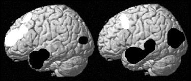
Ictal perfusion changes did not differ between patients with HS and other etiologies. Group 1 had IL anterior temporal lobe hyperperfusion and (pre)frontal lobe hypoperfusion. Group 2 had propagation of IL temporal hyperperfusion towards the posterotemporal regions, and concomitant hypoperfusion of IL posterofrontal and parietal regions (Figure: left= group 1, right=group 2. Black=hyperperfusion, white=hypoperfusion). Group 2 had significant hyperperfusion of the IL thalamus and putamen, and CL parahippocampal gyrus, but not group 1. Group 1 showed weak hypoperfusion (uncorrected p [lt] 0.001) of the CL cerebellum, but not group 2.
Ictal perfusion changes during TL-CPS associated with HS are comparable with the perfusion changes during TL-CPS associated with other etiologies. Posterior propagation of hypoperfusion towards IL posterofrontal and parietal regions concomitant with posterior propagation of ictal temporal hyperperfusion is consistent with the hypothesis that the hypoperfusion represents an ictal surround inhibition. CL cerebellar hypoperfusion occurs during the earlier phase of TL-CPS, while hyperperfusion of IL putamen and thalamus and CL parahippocampal gyrus occurs during a later stage of TL-CPS.[figure1]
[Supported by: Grant Research Fund Katholieke Universiteit Leuven Interdisciplinair Onderzoeksprogramma (IDO) /99/5. ]
