Fluctuating Spike Patterns in Long-Term Intracranial Recordings Among Pediatric Patients
Abstract number :
2.100
Submission category :
3. Neurophysiology / 3G. Computational Analysis & Modeling of EEG
Year :
2018
Submission ID :
502268
Source :
www.aesnet.org
Presentation date :
12/2/2018 4:04:48 PM
Published date :
Nov 5, 2018, 18:00 PM
Authors :
Samuel B. Tomlinson, University of Rochester Medical Center; Camilo Bermudez, Children’s Hospital of Philadelphia; Brenda E. Porter, Stanford Children's Hospital; and Eric Marsh, Children's Hospital of Philadelphia
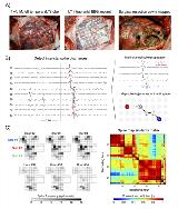
Rationale: Previous studies offer conflicting evidence about the role of interictal spike activity in planning the surgical resection. Studies of multi-day intracranial electroencephalography (IEEG) recordings have documented extensive variability in spike patterns across time, including spike frequency, propagation, and co-localization to the seizure onset zone (SOZ). In this study, we applied novel spike extraction and trajectory clustering tools to characterize the temporal heterogeneity of spike activity in pediatric patients. Methods: Long-term subdural IEEG recordings were retrospectively obtained from 19 pediatric patients (mean age = 11.4 years, range = 3-20 years, mean analyzed recording duration = 54.7 hours). A previously-validated, automated spike detector was used to identify interictal spikes. Propagating spike discharges were extracted using a spatiotemporal clustering algorithm (Fig. 1). The spatial distribution of prominent spike activity was examined across the recording duration, including explicit comparisons of the pre-ictal and post-ictal windows. Spatial spike density maps (spikes/minute/channel) were used to represent spike activity across electroe contacts. Discrete ‘shifts’ in spike patterns were identified using cross-correlation and community partitioning approaches (Fig. 1C). Spike patterns were assessed in relation to time-of-day and seizure proximity. Post-operative outcomes were assessed using the modified Engel outcome scale (minimum =2 years; Class I = ‘Seizure-Free’; Class =II = ‘Seizure-Persistent’). Results: Spike frequency, localization, and propagation trajectories varied tremendously over time (e.g., Fig. 1C for spike density map across time from one representative patient). Across several metrics, Seizure-Free patients exhibited significantly greater temporal stability of spike patterns compared to Seizure-Persistent patients. The relationship between time-of-day and spike frequency was inconsistent across patients. Statistically-significant changes in spike localization and propagation were observed following most seizures; for secondarily generalized seizures, the distribution of spike activity significantly changed in >75% of cases (e.g., Fig. 2 for one representative seizure). On average, spike frequency decreased from pre-ictal to post-ictal windows by 32.1 ± 14.4%, and this effect was driven primarily by secondarily generalized seizures (compared to seizures that did not propagate beyond the SOZ). Conclusions: Spike activity (frequency, localization, propagation, co-localization to the SOZ) is highly variable over time and across patients. On several metrics, temporal instability of spike patterns was associated with less favorable surgical outcomes. Multi-dimensional spike analysis may provide insights into the configuration and stability of epileptic networks prior to surgery. Funding: Funding for this project was provided by the Neurosurgery Research and Education Foundation (NREF). The authors have no conflicts of interest to disclose, commercial or otherwise.

.tmb-.jpg?Culture=en&sfvrsn=9b69ea9f_0)