High-Resolution 3D MR Fingerprinting for Detection and Characterization of Epileptic Lesions
Abstract number :
1.253
Submission category :
5. Neuro Imaging / 5A. Structural Imaging
Year :
2018
Submission ID :
500073
Source :
www.aesnet.org
Presentation date :
12/1/2018 6:00:00 PM
Published date :
Nov 5, 2018, 18:00 PM
Authors :
Z Irene Wang; Stephen Jones, Imaging Institute, Cleveland Clinic; Anagha Deshmane, Magnetic Resonance Center, Max Planck Institute for Biological Cybernetics; Ken Sakaie, Imaging Institute, Cleveland Clinic; Eric Pierre, The Florey Institute of Neuroscien
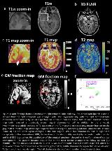
Rationale: Approximately 30% of epilepsy patients are refractory to pharmacotherapy but are potentially curable by surgery. However, conventional MRI can be limited in detecting subtle epileptic lesions or identifying active/epileptic lesions among widespread, multifocal abnormalities. In this study, we developed a high-resolution 3D MRF protocol to simultaneously provide T1, T2, proton density, and tissue fraction maps (gray matter, white matter and cerebrospinal fluid maps) for detection and characterization of epileptic lesions. Methods: A 3D MRF scan with a whole-brain coverage and 1.2 mm isotropic resolution was developed using accelerated MRF acquisition and low-rank model-based reconstruction. This scan simultaneously provided quantitative T1, T2 and proton density maps. Based on the same acquired data, dictionary-based partial volume estimation (PV-MRF) was then performed to generate tissue fraction maps (gray matter, white matter and cerebrospinal fluid maps) that highlight tissue boundaries. Finally, region of interest analysis was performed on T1 and T2 maps to characterize lesions and to differentiate active/non-active lesions. Results: The quantitative T1 and T2 values measured from the International Society for Magnetic Resonance in Medicine/National Institute of Standards and Technology MRI phantom showed high agreement with the standard values. The T1 and T2 maps also showed high repeatability in the volunteer study, with an average coefficient of variance of 3.7% in various normal tissue regions among five volunteers. For the 15 epilepsy patients who were included in the pilot study, MRF showed additional findings in 4 of the 15 patients, with the remaining 11 patients showing findings consistent with conventional MRI. The additional MRF findings were highly concordant with patients’ electroclinical presentation, including invasive evaluation such as stereotactic-EEG. Conclusions: The 3D MRF protocol presented here showed potential to identify otherwise inconspicuous epileptogenic lesions from the patients with negative conventional MRI diagnosis, as well as to correlate with different levels of epileptogenicity when widespread lesions were present. These findings indicate the potential added value of using quantitative MRF protocol as part of presurgical evaluation. Funding: The authors would like to acknowledge funding from Siemens Healthcare and NIH grants NIH 1R01EB016728 and NIH 5R01EB017219.

.tmb-.jpg?Culture=en&sfvrsn=74bb574c_0)