Hippocampal Atrophy in Posttraumatic Epilepsy (PTE)
Abstract number :
2.186
Submission category :
5. Neuro Imaging / 5A. Structural Imaging
Year :
2018
Submission ID :
502629
Source :
www.aesnet.org
Presentation date :
12/2/2018 4:04:48 PM
Published date :
Nov 5, 2018, 18:00 PM
Authors :
Ju-hee Oh, St. Vincent's Hospital, The Catholic University of Korea and Sung Chul Lim, St. Vincent's Hospital, The Catholic University of Korea
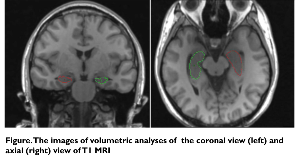
Rationale: It is well known that trauma can produce temporal neocortical damage that in turn produces epilepsy and hippocampal sclerosis frequently coexists with temporal neocortical damage. In a rodent model of traumatic brain injury, the damage in the hippocampal formation is restricted primarily to the hilar region of the dentate gyrus. In several case reports, the loss of hilar neurons in humans may also lead to increased excitability in the hippocampus and subsequently to the development of temporal lobe epilepsy (TLE). We performed a cross-sectional study to determine whether head trauma may induce the secondary hippocampal volume loss. We performed a quantitative MRI volumetry study to compare the hippocampal volume status of our post-traumatic epilepsy (PTE) patients with that of TLE patients who have already had HS. Methods: We reviewed the images and medical records retrospectively. The objects were included who visited or were admitted to St. Mary’s Hospital from February 18th, 2010 to April 30th, 2012. There were 102 patients, however among them we can achieve the data of only 21 patients who had the history of trauma before recurrent seizure without other etiology (post traumatic epilepsy, PTE). Among 21 patients, we could achieve the data in only 8 patients who underwent a T1-volume MRI acquisition. We used manual volumetric analysis using Analyze 10.0 software by one specialist (Jung-Hee Cho) for measuring the volume of hippocampus in each patient. (figure.) We compared the clinical and MRI volumetry data with those of age-matched 10 TLE patients with overt unilateral hippocampal atrophy as control. Results: There was no significant difference between two groups when we compared the average volume of ipsilateral and contralateral hippocampi to seizure focus. The average volume of ipsilateral hippocampi to seizure focus were 2229±405(SD) in TLE patients and 2222±583(SD) in PTE patients (p=0.847). The average volume of contralateral hippocampi to seizure focus were 2561±323(SD) in TLE patients and 2415±255(SD) in PTE patients (p=0.211). (table 1) We saw disproportionate hippocampal atrophy which was more in the ipsilateral hippocampus, less in the contralateral hippocampus, similar to that of control patients with established unilateral hippocampal sclerosis.These findings may support the occurrence of selective neuronal damage in subjects with TLE that develops after head trauma. In a recent study, early electrographic seizures appear to be associated with long-term selective ipsilateral hippocampal atrophy and to a lesser extent contralateral hippocampal atrophy without a net effect on global brain atrophy. A limitation of this study: -Absence of intra-individual follow-up data gives weak causal relationship of each patient’s data -The sample size is small and there is potential for bias in data acquisition and volume measurements. Conclusions: By quantitative MR volumetry study, we could find an evidence of the occurrence of indirect or additional neuronal damage on their hippocampi in PTE patients, . Further studies, such as pathologic change of hippocampus should be warranted to elucidate the mechanism of hippocampal atrophy after traumatic head injury.References1. Swartz BE, Carolyn RH, Tomiyasu U, et al. Hippocampal cell loss in posttraumatic human epilepsy. Epilepsia. 2006;47(8):1373–1382. 2. Vespa PM, McArthur DL, Xu Y, et al. Nonconvulsive seizures after traumatic brain injury are associated with hippocampal atrophy. Neurology. 2010;75:792–798. Funding: None

.tmb-.png?Culture=en&sfvrsn=b290337c_0)