Late Post Hypoxic Myoclonic Epilepsy – Is It Still Lance-Adams Syndrome?
Abstract number :
2.433
Submission category :
18. Case Studies
Year :
2018
Submission ID :
501307
Source :
www.aesnet.org
Presentation date :
12/2/2018 4:04:48 PM
Published date :
Nov 5, 2018, 18:00 PM
Authors :
Ramsha Malik, University of Texas Health Science Center at Houston; Asim Naveed, Baylor College of Medicine; Gerson Suarez Cedeno, University of Texas Health Science Center; and Omotola A. Hope, University of Texas Health Science Center
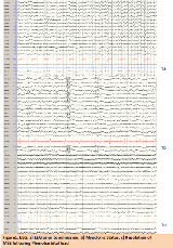
Rationale: Hypoxic injury to the brain may result in Post-Hypoxic Myoclonus (PHM). Acute onset of PHM may be due to Myoclonic Status Epilepticus (MSE) or myoclonus not associated with cortical networks or both. Lance-Adams syndrome (LAS) is the chronic form of PHM and has a delayed onset after the initial hypoxic insult.1 Accurate distinction between the two is important as the prognosis and management is significantly different. We present a case of LAS which challenges our current understanding of MSE and LAS while highlighting a gray area between them. Methods: Case study, literature review Results: 65-year-old male underwent cardiopulmonary arrest during cholecystectomy in March 2017 with return of spontaneous circulation after five minutes. He regained consciousness and had no residual focal deficits, seizure, or evidence of myoclonus. MRI of the brain revealed generalized cerebral volume loss presumed secondary to hypoxic injury. Several weeks later, he was noted to have brief episodes of involuntary twitching of his neck and all four extremities that were triggered by action. Given the characteristics and the late onset of myoclonus, patient was diagnosed with LAS. Myoclonus was managed with Levetiracetam (LEV) and Valproic Acid (VPA).In September 2017, the patient was admitted for a three day history of encephalopathy and increased jerking. Initial workup was negative except hyperammonemia, likely secondary to VPA use. However patient remained encephalopathic despite resolution of hyperammonemia. Routine EEG showed low amplitude polyspike and wave discharges. Treatment with Lacosamide (LCS), LEV, and VPA failed to control his myoclonic seizures and patient remained minimally responsive. Continuous video-EEG revealed higher amplitude and frequency of spike and wave discharges occurring every 2 seconds and correlated with full body jerks. Findings were consistent with MSE. Conclusions: Despite our understanding of the basic differences between MSE and LAS (Table 1), there is significant controversy surrounding the disorders. An overlap may exist, resulting in false diagnosis and inaccurate prognostication. Our patient developed MSE months after being diagnosed with LAS - so with the presence of status myoclonus would this still be considered LAS? Or would the diagnosis change to MSE, a disorder known for its devastating prognosis.2 This case highlights how limited our knowledge of LAS is, perhaps due to the limited number of LAS cases reported in literature. There is need to better define a diagnostic criteria for LAS and MSE, particularly including neurophysiologic characteristics. Origination of myoclonus from cortical versus subcortical areas needs further study, and may correlate with how the myoclonus presents clinically.31. Lance JW, Adams RD. The syndrome of intention or action myoclonus as a sequel to hypoxic encephalopathy. Brain.1963;86:111–136.2. Wijdicks EFM, Hijdra A, Young GB, Bassetti CL, Wiebe S. Practice parameter: Prediction of outcome in comatose survivors after cardiopulmonary resuscitation (an evidence-based review). Neurology..2006;67:203–210.3.Freu nd B, Kaplan PW. Myoclonus after cardiac arrest: Where do we go from here? Epilepsy Currents. 2017;17(5):265-272. Funding: None

.tmb-.png?Culture=en&sfvrsn=d709aa43_0)