Localization of High-Frequency Oscillations with Magnetoencephalography and Electroencephalography Prior to Pediatric Epilepsy Surgery
Abstract number :
1.043
Submission category :
1. Basic Mechanisms / 1C. Electrophysiology/High frequency oscillations
Year :
2018
Submission ID :
502541
Source :
www.aesnet.org
Presentation date :
12/1/2018 6:00:00 PM
Published date :
Nov 5, 2018, 18:00 PM
Authors :
Eleonora Tamilia, Boston Children's Hospital, Harvard Medical School; Matilde Dirodi, Boston Children's Hospital; Steven M. Stufflebeam, Martinos Center for Biomedical Imaging, Massachusetts General Hospital; P Ellen Grant, Harvard Medical School; Joseph
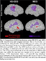
Rationale: For patients with medically refractory epilepsy (MRE), the success of epilepsy surgery depends on the availability of a precise biomarker of epileptogenicity. High-frequency oscillations (HFOs) have emerged as reliable epilepsy biomarkers. Ripple HFOs (>80 Hz) can be seen using magnetoencephalography (MEG) or scalp electroencephalography (EEG), but it is still unknown whether noninvasive techniques localize the HFOs in the same area as intracranial EEG (iEEG) and whether resecting this area predicts outcome. Our goal is to assess the utility of the noninvasive HFO localization prior to epilepsy surgery by: (i) localizing HFOs on MEG and high-density (HD) EEG data from children with MRE; (ii) validating the HFO source localization against the ground-truth HFO-zone defined by iEEG; and (iii) comparing the HFO source localization with resection and outcome Methods: We studied children with MRE who underwent surgery after iEEG. We analyzed simultaneous MEG (306-channels) and HD-EEG (70-channels) data, as well as iEEG data (subdural grids). We detected interictal HFOs (80-160 Hz) on MEG and HD-EEG using an in-house automatic detector followed by the review of an epileptologist, who classified them as with or without spikes. Each HFO was localized on the patient’s cortex using the wavelet Maximum Entropy on the Mean. Only HFOs with spikes were further analyzed, as more likely to be epileptic. We co-registered presurgical magnetic resonance imaging (MRI) with post-implantation computerized tomography (CT) scan to determine the location of each iEEG contact, and with postsurgical MRI to determine the resection. We classified as outside-coverage any HFO localized >15 mm far from iEEG contacts. Then, we estimated the distance of each HFO localization from the ground-truth HFO-zone (defined by iEEG contacts showing HFOs): an HFO was regarded concordant when such distance was <15 mm. Finally, for each patient, we determined whether the HFO-zone localized with MEG or HD-EEG was resected Results: We included 16 children with MRE (11±3.9 years). We found HFOs in all patients using HD-EEG [mean rate: 0.7 HFOs/min (on spikes); 0.5 HFOs/min (no spikes)] and in 13 patients using MEG [mean rate: 0.6 HFOs/min (on spikes); 0.3 HFOs/min (no spikes)]. HD-EEG localized a higher percentage of outside-coverage HFOs (38%) than MEG (20%, ?2: p=0.038, Fig 1). Our data show that: (i) most of outside-coverage HFOs for both HD-EEG (64%) and MEG (56%) were seen in patients with poor outcome (Engel>1b), implying the possibility of an epileptic focus outside the iEEG area (Fig 1), and (ii) most HFOs without spikes were localized outside-coverage (HD-EEG: 57%; MEG: 65%), suggesting their physiological nature. The HFOs localized noninvasively were highly concordant with the ground-truth (Fig 1) for both HD-EEG (92%) and MEG (91%), with no difference between the two modalities (p>0.1). Finally, the HD-EEG HFO-zone was resected in 88% of good-outcome patients (Engel 1a-b), but only in 20% of poor-outcome patients. Similarly, the MEG HFO-zone was resected in 60% of good-outcome patients, but only in 17% of the poor-outcome cases (Fig 2) Conclusions: This study shows that the noninvasive localization of interictal HFOs with MEG and HD-EEG is highly concordant with the ground-truth HFO-zone defined by iEEG in children with MRE. The high percentage of good outcome patients in whom the non-invasively defined HFO-zone was resected, as opposed to the low percentage in poor outcome cases, emphasizes the utility of MEG and HD-EEG to localize HFOs prior to epilepsy surgery, and their possible contribution to guide resection tailoring Funding: None

.tmb-.jpg?Culture=en&sfvrsn=2d0bbfe_0)