Localized, intra-tuberal epileptogenicity in tuberous sclerosis
Abstract number :
3.288
Submission category :
Late Breakers
Year :
2013
Submission ID :
1840533
Source :
www.aesnet.org
Presentation date :
12/7/2013 12:00:00 AM
Published date :
Dec 5, 2013, 06:00 AM
Authors :
A. Harvey C. Bailey R. Hatch, S. Mandelstam, A. Davidson, D. MacGregor, M. Kean, S. Barton, K. Pope, R. Leventer, S. Petrou, W. Maixner
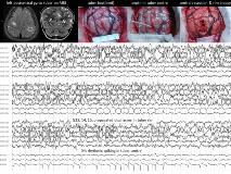
Rationale: We reported intracranial EEG monitoring findings in children with epilepsy and tuberous sclerosis (TS) which suggested tubers are the generators of seizures (Mohamed et al, Neurology 2012;79:2249-57). The frequent observation of trains of rhythmic interictal epileptiform discharges (IEDs) and seizure onsets in the center of epileptogenic tubers led us to investigate in subsequent patients the possibility of epileptogenicity being restricted to the tuber center.Methods: Twelve consecutive children with TS, aged 0.8-18 (median 4) years, underwent epilepsy surgery between April 2012 and September 2013. Sixteen foci were targeted, determined by video-EEG localization of seizures (central 2, frontal 5, temporal 4, parietal 1, occipital 4) and MRI demonstration of candidate tubers (one in 4 foci, multiple in 12 foci). Two patients were operated twice and one patient thrice, for multiple seizure foci. Only one patient underwent intracranial EEG monitoring. All patients underwent (i) preoperative 3T MRI reformatted in planes specific for the candidate tubers, (ii) intraoperative electrocorticography (ECoG) with strip and depth electrodes in, over and around candidate and remote tubers, looking for localized rhythmic IEDs and electrographic seizures, (iii) removal of one or more epileptogenic tubers, based largely on ECoG findings, and (iv) light microscopy of en bloc tuber resections or multiple tuber biopsies. In seven patients, cortical biopsies from epileptogenic tubers were studied in vitro, using 60-contact micro-electrode arrays to record stimulated postsynaptic field potentials. The study was approved by the RCH HREC.Results: MRI revealed a pit or groove in the center of all epileptogenic tubers, with thickened cortex, low T2/high T1 signal and a band traversing the tuber to join the subcortical radial band. Intraoperative ECoG revealed runs of rhythmic IEDs in the center of epileptogenic tubers in 13 surgeries, with (in 7) or without (in 6) propagation to the tuber rim, and independent of any perituberal spiking. Ten patients are free of their targeted seizures (follow-up 1-18 mths), following single (7 foci) or multiple (9 foci) tuberectomies, including 4 patients in whom only the tuber center was resected. Histopathology revealed greater density of dysmorphic cytomegalic neurons in the tuber center compared to the tuber rim. Group analysis of in vitro electrophysiological data revealed significantly different input-output relationships in cortical slices from 4 tuber centers compared to 7 tuber rims.Conclusions: The localized radiological, histological and electrical abnormalities in the center of epileptogenic tubers, and the control of seizures with resection of the tuber center only, suggest epileptogenicity in TS is due to a focal cortical dysplasia with dysmorphic cytomegalic neurons in the center of an otherwise 'inert' tuber. A simple approach to epilepsy surgery may be possible in some children with TS, localizing epileptogenic tubers with video-EEG, MRI and ECoG, and potentially confining resection to the tuber center. Dr Hatch was supported by a grant from the Royal Children's Hospital Foundation.
