Multicenter Harmonization of Preclinical MRI Biomarker Discovery in a Rat Model of Post-Traumatic Epileptogenesis
Abstract number :
1.097
Submission category :
2. Translational Research / 2C. Biomarkers
Year :
2018
Submission ID :
501356
Source :
www.aesnet.org
Presentation date :
12/1/2018 6:00:00 PM
Published date :
Nov 5, 2018, 18:00 PM
Authors :
Riikka J. Immonen, University of Eastern Finland; Gregory Smith, David Geffen School of Medicine at UCLA; Rhys D. Brady, Central Clinical School, Monash University; David Wright, Central Clinical School, Monash University; Leigh A. Johnston, The Royal Mel
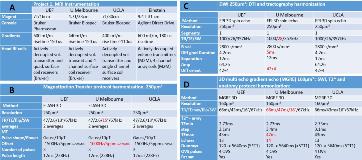
Rationale: Preclinical imaging studies of post-traumatic epilepsy (PTE) have largely been proof-of-concept studies with limited animal numbers, and thus have often lacked the statistical power for biomarker discovery. The Epilepsy Bioinformatics Study for Antiepileptogenic Therapy (EpiBioS4Rx) is a pioneering multicenter study of preclinical imaging biomarkers of PTE that was faced with the need to harmonize the MRI procedures and data metrics prior to its execution. We present here the harmonization process and evaluate the uniformity of the obtained MRI data. Methods: The consortium includes four preclinical MRI facilities at University of Eastern Finland (site 1), University of Melbourne/Monash University (site 2), University of California at Los Angeles (site 3), and Albert Einstein College of Medicine (site 4) hosting magnets of 7T, 4.7T, 7T, and 9.4T field strengths, respectively. Sites 1-3 participated this first phase. MRI techniques compatible with each hardware, including 3D diffusion tensor imaging and tractography, magnetization transfer imaging, multi echo gradient echo - and T2wt-sequences, were optimized for probing TBI pathology (lateral fluid percussion injury in Sprague Dawley adult male rats) and tailored to produce uniform quantitative data across different field strengths (Table1). MRI was repeated 2, 9, and 30 days post injury. Data from sham operated rats (Ntotal=24; N=8 from each site) were analyzed and MRI signal uniformity between sites (at 30d), signal variation in combined cohort, repeatability over time, and variation in timing of MRI scans were assessed. Quality control scans using phantoms were executed prior to each batch to monitor hardware stability. Results: Process. Four iterations of optimization and feedback, shared methods, and distributed instructions yielded successful harmonization of MRI data. Signal. Signal to noise ratios (SNR) in T2wt images were 34.9±2.2 (site1), 47.7±2.2 (site2), and 32.1±1.7 (site3), with a pooled average of 37.7±7.0. Magnetization transfer ratios (MTR) were 58.4±0.5% (site1), 56.5±0.6% (site2), and 57.6±1.1% (pooled average) in gray matter, and 67.0±0.7% (site1), 65.3±0.1% (site2) and 66.2±1.2% (pooled average) in white matter (WM). Thus, the GM/WM MTR contrast was uniform between sites (p>0.05). Fractional anisotropy in WM was 0.80±0.02, 0.79±0.03 and 0.79±0.04 in sites 1-3, respectively. Repeatability. Uniform SNRs were obtained between the repeated scans in site 1 and 2. SNR in site 3 improved from 9 to 30 days post operation. Time of scan from the injury was 48.0±0.11 hours, 216.0±0.12 hours and 30.0±2.2 days (site1, N=56), and 55.4±10.9 hours, 228.1±33.7 hours and 28.7±1.9 days (site3, N=24), p<0.05 at day 2. QC phantom scans demonstrated hardware performance to be stable over time (data not shown). Conclusions: MR signal properties were harmonized with only modest variation between sites despite utilizing scanners of different field strength, allowing pooling data from all sites. Deviations in timing of the acute scans are to be taken into account as a covariate. Funding: Supported by NINDS Center without Walls, U54 NS100064 (EpiBioS4Rx)

.tmb-.jpg?Culture=en&sfvrsn=4b4a6ff4_0)