Photoplethysmography: A Measure for the Function of the Autonomic Nervous System During Focal Impaired Awareness Seizures
Abstract number :
3.095
Submission category :
2. Translational Research / 2C. Biomarkers
Year :
2018
Submission ID :
506363
Source :
www.aesnet.org
Presentation date :
12/3/2018 1:55:12 PM
Published date :
Nov 5, 2018, 18:00 PM
Authors :
Bethany Bucciarelli, Boston Children's Hospital, Harvard Medical School; Rima El Atrache, Boston Children's Hospital, Harvard Medical School; Eleonora Tamilia, Boston Children's Hospital, Harvard Medical School; Sarah Hammond, Boston Children's Hospital,
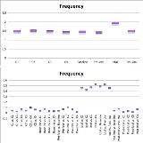
Rationale: Photoplethysmography (PPG) uses reflected light waves to detect volumetric changes in arterial blood flow. By reflecting variations of blood perfusion in tissues, PPG may allow the detection of changes in autonomic nervous system (ANS) function. The aim of this study is to assess the variability of PPG signals and their value in detection of peri-ictal changes in ANS function in patients with focal impaired awareness seizures, formerly known as complex partial seizures (CPS). Methods: PPG data was recorded using a wristband sensor (E4, Empatica Inc., Milan, Italy) from patients undergoing long term video EEG monitoring. Patients’ EEG and seizure videos served as the gold standard to identify seizure onset and offset times as well as their seizure types. PPG signals were analyzed in four timeframes: baseline, preictal, ictal and postictal. Four control portions of signals were extracted per patient from seizure free days. We used MATLAB to extract features from the PPG signal, including frequency, duration, amplitude, decreasing and increasing slope, smoothness, and area under the curve (AUC). The analysis was performed using a linear mixed-effects model to account for within subject correlations Results: We enrolled 213 patients from the long term video EEG monitoring unit at Boston Children’s Hospital between February 2015 and January 2018. The age of patients ranged from 7 to 15 years. We excluded seizures that were not clearly focal impaired awareness seizures, when the patient was out of view of the video camera, those that occurred within 90 minutes or less of other seizures, and those with PPG signals affected by major motion artifacts. As a result, we included 11 patients with a total of 18 focal impaired awareness seizures. The ictal phase was excluded from 6 seizures (4 patients) as it was affected by extensive motion artifacts.PPG frequency increased significantly during the ictal period with an average increase of 0.46Hz (p<0.0001), compared to an average variability of 0.04Hz during the control periods. The average increase in frequency was 0.48Hz when compared to the control, baseline and pre-ictal phases together.Duration was different in the ictal phase with an average of 0.18s (p<0.0001), compared to the control with an average change of 0.02s. When comparing ictal duration to the control, baseline and pre-ictal phases combined the average duration is 0.19s. Conclusions: PPG signals change during the ictal phase in patients with focal impaired awareness seizures. PPG sensors may serve as a marker for detection of this type of seizure. Further work is needed to better understand the benefit of employing PPG in detection of focal impaired awareness seizures (Funded by the Epilepsy Research Fund). Funding: Epilepsy Research Fund

.tmb-.jpg?Culture=en&sfvrsn=adfd525e_0)