Preictal high-frequency oscillations in scalp EEG are localized in the brain region of seizure onset
Abstract number :
3.309
Submission category :
Late Breakers
Year :
2013
Submission ID :
1861665
Source :
www.aesnet.org
Presentation date :
12/7/2013 12:00:00 AM
Published date :
Dec 5, 2013, 06:00 AM
Authors :
C. Stamoulis, , B. Chang
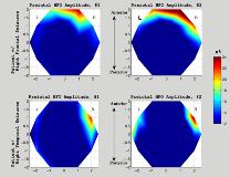
Rationale: Transient, low-amplitude high-frequency oscillations (>80 Hz) have recently been estimated in scalp EEG from patients with focal epilepsy, at least a few minutes prior to ictal onset. For these oscillations to be measurable at the scalp they must originate from relatively large areas of the brain (at least 6 cm2 of cortex), i.e., beyond the epileptogenic region. It is unclear whether these oscillations are spatially distributed across the entire brain or are correlated with the region of seizure onset. Methods: Scalp EEGs from 9 patients (age 26-50 years) with diagnosed focal epilepsy, which included preictal intervals at least 2 minutes prior to ictal onset, were analyzed. Signals were recorded with an International 10-20 system and were sampled at 500 samples/sec. Based on visual examination of the EEG, 3 patients had frontal seizures and 6 patients had temporal lobe seizures. EEGs were first pre-processed to suppress artifacts and were then decomposed using developed computational techniques for estimating non-random high-frequency waveforms in scalp recordings. Both the characteristic frequency and peak amplitude of these waveforms were estimated. A total of 52 preictal segments (2-10 min long) were analyzed.Results: In patients with seizures originating in right temporal lobes, the highest preictal HFO amplitude was consistently estimated in temporal/fronto-temporal regions. Figure 1 (bottom panels) shows two examples of spatial HFO distribution estimated from preictal intervals in one patient with right temporal seizures. HFO amplitude was averaged over time. In both examples, the highest HFO amplitude was estimated in channels F8 and T2. Characteristic HFO frequencies were in the range 132-158 Hz. In patients with frontal seizures, higher spatial variability of HFO amplitude and frequency was estimated. In some preictal intervals the highest preictal HFO amplitude was detected in either right or frontal regions, in agreement with the visual EEG examination. However, in a few preictal intervals high-amplitude HFOs were detected bilaterally or in larger brain areas. Figure 1 (top panels) shows two examples of spatial HFO distribution estimated from preictal intervals in one patient with right frontal seizures. In the left panel, HFO amplitude was highest in frontal channels Fp2, F8 and F4. In the right panel high HFO amplitude was estimated bilaterally, and extended to right fronto-temporal regions. HFO frequencies were in the range 147-176 Hz.Conclusions: HFOs in preictal scalp EEG appear to be spatially localized in the brain region of seizure onset, or broader areas that include this region. In patients with temporal lobe seizures, there was a consistent correlation between the origin of seizure onset identified by visual examination of the EEG and peak HFO amplitude. In patients with frontal seizures, HFOs were localized either in the lobe of seizure onset, or in some cases bilaterally or broader regions. These results suggest that preictal HFOs are correlated with the region of seizure onset and are not spatially distributed across the entire brain.
