Rapid Improvement of Encephalopathy and EEG in Children With Lennox-Gastaut Syndrome and Generalized Epilepsy After Focal Resection
Abstract number :
3.343
Submission category :
9. Surgery / 9B. Pediatrics
Year :
2018
Submission ID :
502611
Source :
www.aesnet.org
Presentation date :
12/3/2018 1:55:12 PM
Published date :
Nov 5, 2018, 18:00 PM
Authors :
Akshat Katyayan, Boston Children's Hospital and Masanori Takeoka, Boston Children's Hospital
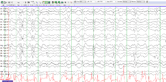
Rationale: Successful epilepsy surgery in children with generalized EEG findings but with focal lesions on neuroimaging has been well- described (by Willie E et al, 2007), with good long term seizure outcome (up to 72% seizure free). However, data is scarce on immediate clinical and EEG improvement in such children. This may be important for children with epileptic encephalopathies, such as Lennox-Gastaut syndrome (LGS), where improvement in encephalopathy and neurocognitive comorbidities are equally important Methods: We describe 2 children who had clinical care for medically intractable epilepsy at Boston Children’s Hospital, who met criteria for LGS (multiple seizure types, including tonic seizures, slow spike wave complexes on EEG and developmental delay) and had focal lesions on neuroimaging. Results: First case was a 3 year old boy who developed epilepsy at 15 months, including epileptic spasms, atypical absence, generalized tonic clonic and myoclonic-tonic seizures. He developed significant language and behavioral regression with onset of epilepsy. He was diagnosed with autism and developed significant behavioral dysregulation. MRI showed a left fusiform gyrus low grade neoplasm. EEG showed abundant multifocal and generalized spike and poly-spike and wave complexes (figure 1), and continuous high amplitude slowing. He became seizure free immediately after resection of the lesion, and had improvement in his encephalopathy (improved attention span and behavioral regulation). EEG one week after surgery showed normal voltage, noticeable posterior dominant rhythm (figure 2), and sleep spindles. Multifocal and generalized spike waves were seen in isolation, markedly less compared to before surgery. Second case was a 6 year old girl who had normal development until age 3, including talking in full sentences, when she developed tonic seizures, with bilateral upper extremity extension and trunk flexion occurring multiple times a week. She developed neuro-cognitive regression after epilepsy onset, became non-verbal, and lost her skills of daily living. EEG showed multifocal spikes, generalized slow spike wave complexes and generalized tonic seizures. MRI showed a right temporal neoplasm. She underwent resection of the lesion, and became seizure free immediately after surgery. Within one month of surgery, she was able to speak a few words, and started speaking in phrases by one year after surgery. She was able to follow basic instructions immediately after surgery and regained some independence in her activities of daily living. EEG obtained 4 months after surgery showed normal background activity and rare right central and left temporal spikes. Conclusions: Epilepsy surgery in children with epileptic encephalopathies such as LGS with focal lesions on neuro-imaging may not only result in good seizure control but may also lead to early and rapid improvement in clinical encephalopathy, which may be corroborated on EEG. Larger scale studies are needed to further explore the reproducibility of findings.ReferenceWyllie E et al. Successful surgery for epilepsy due to early brain lesions despite generalized EEG findings. Neurology. 2007; 69:389-397. Funding: None

.tmb-.png?Culture=en&sfvrsn=86872dbf_0)