Real-Time Detection of After-Discharges Improves Electrical Brain Stimulation Mapping
Abstract number :
3.151
Submission category :
3. Neurophysiology / 3E. Brain Stimulation
Year :
2018
Submission ID :
506485
Source :
www.aesnet.org
Presentation date :
12/3/2018 1:55:12 PM
Published date :
Nov 5, 2018, 18:00 PM
Authors :
Christoph Kapeller, Guger Techologies OG; Johannes Grünwald, Guger Techologies OG; Kyousuke Kamada, Asahikawa Medical University; and Christoph Guger, Guger Techologies OG
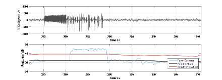
Rationale: After-Discharges (ADs) are electroencephalographic (EEG) seizure patterns following electrical brain stimulation (ECS). Such waveforms indicate that continued stimulation or increased current may provoke epileptic seizures. Termination of AD by follow-up brief bursts of pulsed stimulation can reduce the number of ADs, potentially reduce the risk of seizures or even abort them. One of the difficulties in a clinical stimulation setting is the manual monitoring of hundred or more intracranial EEG channel during functional brain mapping via ECS, which is mandatory in the course of neurosurgical treatment. Hence, automated AD detection simplifies their monitoring and speeds up their localization. Methods: This study introduces a detector that is based upon the principle that ADs usually exhibit tremendously higher signal power than regular EEG signals. The detector computes a short-term power estimate in time domain and compares it to a long-term power estimate that reflects the average EEG signal power. This long-term power estimate is only updated in absence of high-power interference, such as from motion or ECS artifacts. Once the short-term power exceeds mean + k*variance of the long-term power (where k is adjustable), the respective signal segment is considered abnormal. Since AD are causally related to ECS, an identified segment of high power is only considered an AD if it appears shortly (i.e., within 10 s) after the stimulation. Otherwise it is classified as a generic suspicious waveform and can be used for signal quality checks. Incoming EEG data are real-time processed in a sliding-window approach, for each channel individually, where a window length of one second and a step size of 100 ms yielded good results. Feedback about occurring ADs and their location is provided instantaneously to the operator.In this work, we evaluated data from ECS sessions conducted with two patients that suffered from intractable epilepsy and underwent surgical treatment. During ECS, bipolar and biphasic electrical stimulation pulses of 200 µs were injected in 2-5 s trains of 50 Hz with a maximum current of 6 mA for motor and 12 mA for language mapping. The number of channels were 188 for Patient 1 (P1) and 60 Patient 2 (P2). We evaluated 15 stimulation events for P1 and 13 stimulation events for P2. For both Patients, ADs in 266 channels related to the stimulation events were found via visual inspection. In other words, ADs were present in 7.4 % of all channels. Two of five known AD classes (Blume et al., 2004) were found in the data. Results: The proposed detector identified 82 % of all channels containing ADs. No false positives were generated. From the channel-based detector results, an AD was considered globally present if it was detected in at least one channel (on average, 13.3 channels were affected). On a global scope, perfect performance of AD detection (zero false positives and zero false negatives) was achieved. Conclusions: The prompt response of the detector supports the operator of a stimulation mapping to efficiently localize ADs and terminate them by follow-up brief bursts of pulsed stimulation. Such a follow-up stimulation could be even initiated automatically to minimize the number of ADs in a mapping session. It remains to gather and validate the detector with datasets that contain all five AD classes. Funding: This work was supported by the European Union Eurostars project RAPIDMAPS 2020 (ID 9273) and in part by a Japanese Grant-in-Aid for Scientific Research (B) No. 16H05434 from 2016 to 2019.
