THE PATIENT WITH COMPLEX PARTIAL EPILEPSY OF TEMPORAL LOBE ORIGIN REVEALS UNIQUE HEAD SURFACE GEOMETRY OF BASAL TEMPORAL INTERICTAL EEG TRANSIENTS (BTIET)
Abstract number :
1.095
Submission category :
Year :
2002
Submission ID :
3587
Source :
www.aesnet.org
Presentation date :
12/7/2002 12:00:00 AM
Published date :
Dec 1, 2002, 06:00 AM
Authors :
Fumisuke Matsuo, Tawnya M. Constantino, Lorie Blair. Department of Neurology, University of Utah School of Medicine, Salt Lake City, UT
RATIONALE: BTIET can reliably predict complex partial epilepsy, but their accuracy in localizing the epileptogenic neuronal matrix is questioned. A recent preliminary study suggested that BTIET geometry did not differentiate patients with mesial temporal sclerosis (MTS) from those without, as demonstrated by high resolution MRI. This study was designed to evaluate the range of variation in BTIET geometry within each patient and between patients.
OBJECTIVE: The material presented will enable the viewer to understand the principle of localization of EEG epileptiform discharges.
METHODS: Digital EEG data were available from a total of 20 patients, and all the BTIET were identified. BTIET were reformatted in polygraphic display in serial 10-20 bipolar derivations, consisting of anterior-posterior and basal chains (J. Clin. Neurophysiol. 3[Suppl 1]: 26-33). Time series display gain was set to equalize the height of the primary BTIET peak. The time axis of BTIET display was graphically adjusted so that the primary BTIET peak and one immediately following it could be superposed. Geometric variation could be readily appreciated in display, superposing a varying number of BTIET.
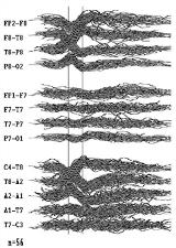
RESULTS: A total of 937 BTIET were collected (individual patients contributing one to 426 BTIET). Despite substantial variation in waveform among BTIET, superposition indicated a unique localization of BTIET source in each patient. Superposed BTIET could differentiate different patients, based on subtle but definite variation in voltage gradient over the head surface. Figure illustrates a total of 56 BTIET superposed from a patient. Instrumental phase reversal indicates middle temporal source localization. [figure 1]
CONCLUSIONS: Head surface EEG geometry derived from 10-20 head surface electrode placements diffentiated source localization of BTIET in this group of patients. Subregional localization of the epileptogenic matrix thus accomplished may offer a simple, noninvasive clinical tool for improved clinical neuroanatomical correlation. Further investigations are warranted to determine whether BTIET geometry can differentiate patients with MTS from those without and between primary and secondary BTIET.
