TIME COURSE OF QUANTITATIVE EEG CHANGES DURING CARDIAC ARREST AND RESUSCITATION
Abstract number :
2.401
Submission category :
Year :
2014
Submission ID :
1868953
Source :
www.aesnet.org
Presentation date :
12/6/2014 12:00:00 AM
Published date :
Dec 4, 2014, 06:00 AM
Authors :
Babak Razavi and Kimford Meador
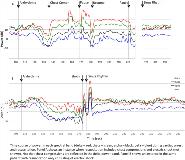
Rationale: Scalp electroencephalography (EEG) can be used as a surrogate marker for cerebral perfusion in a variety situations including cardiac arrest. Prior reports have demonstrated qualitative EEG changes with cerebral hypoperfusion related to cardiac arrest and subsequent reperfusion with resuscitation. However, to our knowledge the time-course of EEG changes in the setting of cardiac arrest has not been described using quantitative methods. We describe quantitative spectral analysis of EEG in a patient undergoing cardiac resuscitation. Methods: The patient is a 60 year-old male who was admitted to the intensive care unit after suffering cardiac arrest. He was placed on cooling protocol and mechanical ventilation, and continuous scalp EEG recording (200 Hz sampling rate) using standard 10-20 electrode locations. He had 3 separate episodes of ventricular arrhythmia, for which he underwent resuscitation with chest compressions, electric shock, and epinephrine. Scalp EEG recordings were quantified by calculating the power in each spectral band (delta, theta, alpha, beta) at each electrode location (bipolar double banana montage) over short-duration sliding time windows with partial overlap. Results: After onset of ventricular arrhythmia, changes in spectral band powers emerged as early as ~10 seconds across all spectral bands, when subtle changes may be appreciated on EEG by visual inspection. The power for each spectral band eventually decreased, then reached a minimum, corresponding to EEG tracing showing minimal activity. During this time power remained higher in the slower frequency bands (delta and theta) than the faster ones (alpha and beta). Chest compressions were associated with re-emergence of EEG activity including faster frequencies. Pauses in chest compressions resulted in recurrent loss power across frequencies. After resumption of sinus rhythm, power in all frequency bands recovered fairly quickly though more gradually for the faster frequencies (alpha and beta). Conclusions: We have used spectral techniques to describe the time course of EEG changes related to cardiac arrest. These were detected when EEG changes are subtle on visual inspection. Our analysis demonstrates that quantitative EEG is a powerful surrogate marker for cerebral perfusion during compromised cardiac function. This case underscores the importance of immediate and effective resuscitation including chest compressions to maintain perfusion and minimize cerebral injury.
