Two-Step Combined Stereo-EEG and Chronic Subdural EEG Recording for Epilepsy Surgery
Abstract number :
2.304
Submission category :
9. Surgery / 9A. Adult
Year :
2018
Submission ID :
501421
Source :
www.aesnet.org
Presentation date :
12/2/2018 4:04:48 PM
Published date :
Nov 5, 2018, 18:00 PM
Authors :
Hideaki Tanaka, Fukuoka Sanno Hospital; Hiroshi Shigeto, Fukuoka Sanno Hospital; Shinji Ohara, Fukuoka Sanno Hospital; Tooru Inoue, Fukuoka University Hospital; and Naoki Akamatsu, International University of Health and Welfare
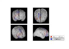
Rationale: Both Stereo-EEG (SEEG) and chronic subdural EEG recording are important tools for estimation of the epileptogenic zone (EZ). Each method has advantages and disadvantages; SEEG enables to investigate the deep structure and distant area with fewer stresses compare to subdural EEG recording, while functional mapping and cortical surface information are highly limited. Methodology to utilize the advantages of each method is highly recommended. We aim to demonstrate two-step method using SEEG and subdural EEG recording in a case with unclarified EZ based on non-invasive studies. Methods: As a first step, we insert small number of SEEG using frame-based stereotactic apparatuses based on the information of non-invasive studies. Scalp electrodes were also attached as many as possible. We roughly estimate EZ based on the findings of SEEG and scalp EEG recording. As second step, chronic subdural EEG recording was performed. The location of subdural electrodes placement was decided based on the findings of the SEEG recording. For subdural EEG recording, we used the grids and strips electrodes. The locations of SEEG and subdural electrodes were confirmed using post-implantation CT superimposed on pre-implantation MRI. Results: A patient was a 21-year-old male with refractory focal epilepsy. MRI showed no any lesion. During long-term video scalp EEG, the patient showed bilateral tonic posture followed by hyper motor seizure. Ictal EEG showed irregular theta activities arising from wide spread anterior part with little deviation to the right. We presumed the EZ was in the right or left frontal lobe, however we needed more information to estimate EZ; medial or lateral frontal lobe, right or left hemisphere, including temporal lobe involvement. Firstly, we selected the SEEG method to find approximate EZ. Four electrodes were implanted using frame device, and targets of each electrode were right orbitofrontal, right temporo-polar, right medial frontal, and left medial frontal lobes. Seizure-onset zone (SOZ) was found in the right inferior frontal area including orbitofrontal gyrus and medial frontal lobe synchronously (Figure 1). We assumed that this patient had the wide EZ in right frontal lobe, and functional mapping was needed. Secondly, after 3 months of SEEG recoding, subdural electrodes were implanted. Eight subdural electrodes and 85 contacts were implanted over the right medial, lateral, and orbitofrontal areas. SOZ was identified in the right middle to inferior frontal and medial frontal gyrus synchronously as figure 2. In cortical stimulation for functional mapping, presumed EZ was not included the eloquent area. Finally, we performed cortical resection of right frontal areas including SOZ and irritative zone. This patient sustains seizure free for one year after resection. Conclusions: We introduced two-step method combined SEEG and subdural EEG. This method is useful for the patient with non-lesional epilepsy with unclarified EZ based on non-invasive studies. We believe two step method may improve the estimation of the EZ more accurately and less invasively. Funding: None

.tmb-.jpg?Culture=en&sfvrsn=699ce8e3_0)