Unprecedented Manifestation of Multisensory Hallucinations in Epilepsy
Abstract number :
3.440
Submission category :
18. Case Studies
Year :
2018
Submission ID :
502691
Source :
www.aesnet.org
Presentation date :
12/3/2018 1:55:12 PM
Published date :
Nov 5, 2018, 18:00 PM
Authors :
Mark Rosenberg, Universidad Autonoma de Guadalajara School of Medicine; Hannia Ramos Salinas, Universidad Autonoma de Guadalajara School of Medicine; and Hanul Bhandari, Nidraveda Neurology and Sleep Medicine Specialists
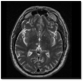
Rationale: Mr. G is a 65-year-old Caucasian male who presented with altered mental status and ataxia. He had confusion to time, place, and situation and upon standing fell to the left side that lasted 30 minutes. While in the ED the patient suffered a sequence of symptoms over several hours, each episode spontaneously resolving after several seconds, including: witnessing a black cat enter his room, momentary confusion and disorientation, holding, tasting and visualizing/”enjoying” a chocolate peppermint candy, and hearing his infant niece crying for about 30 seconds. After being admitted, the patient suffered another episode of seeing a similar black dog enter the room several seconds in duration. Lastly, while urinating he lost balance, fell forward, and then quickly regained balance and returned to his bed. Methods: We theorize that the patient developed a nidus in the temporal lobe, which propagated through the amygdala and disseminated through the thalamus, involving the ventral posterior nucleus (VPN), ventral posterolateral nucleus (VPL), medial geniculate nucleus (MGN) and lateral geniculate nucleus (LGN). By activating the amygdala, the patient’s emotions fluctuated, indicated by the happiness in eating the candy. Furthermore, stimulation of each individual thalamic sensory nuclei is suggested by the separate sensory hallucinations. By affecting the different thalamic relay points, faulty signals were transmitted back to the cerebellum, parietal lobe, temporal lobe and occipital lobes (accounting for the ataxia, gustatory/auditory hallucinations and visual hallucinations, respectively). Results: MRI was performed demonstrating a 9.6 mm acute lacunar infarct in the right thalamus and moderate small vessel disease in the white matter throughout the cerebral hemispheres. Long-term EEG indicated several sharp spike and wave patterns isolated to the temporal field. Conclusions: This patient presents as the first documented case of tactile and gustatory hallucinations connected to epilepsy as well as demonstrating very uncommon and extreme manifestations of auditory and visual hallucinations, suggesting a wider range of intensity of epileptiform hallucinations than previously seen. Funding: No funding was received.
