Very High Frequency Oscillations Using Magnetoencephalography in Patients With Epilepsy
Abstract number :
2.053
Submission category :
3. Neurophysiology / 3D. MEG
Year :
2018
Submission ID :
501390
Source :
www.aesnet.org
Presentation date :
12/2/2018 4:04:48 PM
Published date :
Nov 5, 2018, 18:00 PM
Authors :
Tao Yu, Beijing Institute of Functional Neurosurgery, Xuanwu Hospital, Capital Medical University; Fan Wang, State Key Laboratory of Brain and Cognitive Science, Chinese Academy of Sciences; Xueyuan Wang, Beijing Institute of Functional Neurosurgery, Xuan
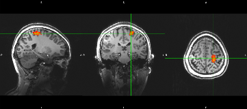
Rationale: High frequency oscillations (HFOs) including ripples (80-200Hz) and fast ripples (200-500 Hz) recorded from intracranial electrodes is a promising biomarker for epilepsy. The localization of very high frequency oscillations (VHFOs) (1000-2000 Hz) was testified more focal and closer to the epileptogenic zone comparing with traditional HFOs in some recent reports. On the other hand, how to get the HFOs information noninvasively in the presurgical evaluation is attractive work. Some recent studies showed that the HFOs could also be recorded by magnetoencephalography (MEG) and these HFOs might be helpful to localize the epileptogenic zone.The aim of this study is to evaluate the probability of localizing the epileptogenic zone using the source localization of VHFOs recorded by MEG. Methods: Three patients with refractory epilepsy were included in this study. These patients underwent presurgical evaluation in Xuanwu Hospital, Beijing. Additionally, MEG data was collected for these patients in a CTF-MISL 275 channel MEG system with sampling rate of 19200 Hz in Institute of Biophysics, Chinese Academy of Sciences, Beijing. The raw data was processed for noise reduction and resampling to 4800 Hz. For HFOs between 200-500 Hz, the raw data was filtered and searched in the time-frequency domain using wavelet transform to locate the peak HFOs in a 30min scan. 10Hz bandwidth around the peak frequency and 1.5s time course were selected to perform source localization use accumulate source imaging in MEG processor. In the 1000-2000 Hz, VHFOs were located using the same way while eliminating the multiplier frequencies of HFOs below 1000Hz. Consecutive MRI images were collected in Siemens 7T system using MP2RAGE. Results: In all three patients, the source localization of HFOs (200-500Hz) by MEG using ASL showed that the HFOs were well co-located in frontal lobes or temporal lobes with the lesion. However, in occipital lobe and cerebellum, there were also some HFOs. The activations may be related to the physiological HFOs involved in the motor system. On the contrary, the VHFOs were only located in bilateral frontal lobes. The source amplitudes in the lesion side were larger than that in the contralateral mirror side. In the time-frequency spectrogram, VHFOs appears to be narrow band with very short lasting time. The VHFOs were more focal with a smaller FWHM than the HFOs. The lesion was further confirmed with stereoelectroencephalography (SEEG). After resection of the epileptogenic zone in this area, the patients acquired seizure free for more than 12 months. Conclusions: In this study, we have demonstrated that VHFOs could also be localized noninvasively in MEG using ASL, and the VHFOs are more focal than HFOs. Combination of HFOs and VHFOs located by MEG might offer more useful information about the epileptogenic zones and the mechanism underlying epilepsy. Funding: This study was funded by National Natural Science Foundation of China [grant numbers: 81771395].
