Two Different Semiology Patterns and Epilepsy Networks of FCD Located in the Superior Frontal Sulcus
Abstract number :
1.017
Submission category :
1. Basic Mechanisms / 1A. Epileptogenesis of acquired epilepsies
Year :
2018
Submission ID :
502060
Source :
www.aesnet.org
Presentation date :
12/1/2018 6:00:00 PM
Published date :
Nov 5, 2018, 18:00 PM
Authors :
Chao Zhang, Beijing Tiantan Hospital, Capital Medical University; Wen-han Hu, Beijing Neurosurgical Institute, Capital Medical University; Xiu Wang, Beijing Tiantan Hospital, Capital Medical University; Bao-tian Zhao, Beijing Tiantan Hospital, Capital Med
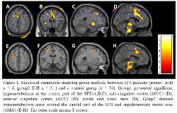
Rationale: Focal cortical dysplasia (FCD) is a common histopathological substrate of focal epilepsy, which tends to occur in the depth of sulci, for instance, the superior frontal sulcus(SFS). The Main purpose of this study was to describe the clinical phenotype, functional metabolic characteristics, and epilepsy network of focal epilepsy with FCD located in the superior frontal sulcus. Methods: Clinical features from 13 Patients with FCD located in the SFS were retrospectively analyzed. The SFS were identified visually and automatically by brain recognition software (Brainvisa) to ascertain the anatomical relationship between the lesions and SFS. Then, the interictal PET data of patients were compared with those of 54 healthy controls using statistical parametric mapping (SPM) to identify regions with significant hypometabolism. For seven patients underwent stereotactic electroencephalography (SEEG), the seizure onset localization and connected neuronal networks were analyzed visually and by two quantitative approaches (‘Epileptogenicity Index’ and ‘Epileptogenicity Maps’). Results: All the patients were seizure free post-operatively. Two distinctive semiology subgroups were identified according to the video EEG analyzing. For Group 1, the patents showed onset semiology of fear/pouting/Hypermotor, and however, for Group 2, the semiology was characterized by versive/limbs tonic and tonic. Compared to controls, Group1 presented significant hypometabolism in the rostral part of the SFS, mid-cingulate cortex (MCC), anterior cingulate cortex (ACC), insula and brain stem, Group 2 showed hypometabolism areas around the caudal part of the SFS and supplementary motor area (SMA). The intracranial EEG analysis results were parallel the SPM findings. In Group 1, 4 SEEG patients showed an early involvement of the MCC and ACC, on the other hand, 3 SEEG patients from Group2 showed the early propagation of SMA with no ACC activation. Conclusions: The epilepsy origin of SFS can be manifested as two types of symptoms. One type of epilepsy originates from the rostral part, and the other one came from the caudal part. The distinctive difference of functional imaging and epilepsy networks may account for the manifestation of semiology. Patients with rostral SFS lesion need to be differentially diagnosed with medial and basal frontal epilepsy. Funding: Capital’s Funds for Health Improvement and Research (CFH, 2016-1-1071); Hainan Province Special Funds for Application Technology Research, Development, and Popularization (ZDXM2015068); Capital Medical University Basic and Clinical Cooperative Research Project (17JL05); Beijing Municipal Party Committee Organization Department Talents Project (2016000021469G214); CAAE UCB Fund Grant (2017002)

.tmb-.jpg?Culture=en&sfvrsn=b77e631a_0)